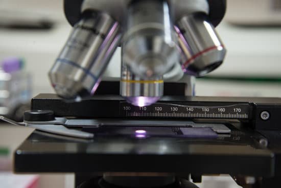
What types can be studied with an electron microscope? Electron microscopes are used to investigate the ultrastructure of a wide range of biological and inorganic specimens including microorganisms, cells, large molecules, biopsy samples, metals, and crystals. Industrially, electron microscopes are often used for quality control and failure analysis.
Was the microscope invented in the 17th century? After its invention in the 1620s, the microscope had its first high point in the second half of the 17th century.
What age was the microscope invented in? A Dutch father-son team named Hans and Zacharias Janssen invented the first so-called compound microscope in the late 16th century when they discovered that, if they put a lens at the top and bottom of a tube and looked through it, objects on the other end became magnified.
Who invented the microscope in 1666? Antoni Van Leeuwenhoek (1635-1723) was a Dutch tradesman who became interested in microscopy while on a visit to London in 1666. Returning home, he began making simple microscopes of the sort that Robert Hooke had described in his, Micrographia, and using them to discover objects invisible to the naked eye.
What types can be studied with an electron microscope? – Related Questions
How robert hooke developed his compound microscope?
To combat dark specimen images, Hooke designed an ingenious method of concentrating light on his specimens, as shown in the illustration. He passed light generated from an oil lamp through a water-filled glass flask to diffuse the light and provide a more even and intense illumination for the samples.
How do microscopic animals eat?
They feed directly on phytoplankton, bacteria and other protozoa. Their respiration releases much of the carbon dioxide incorporated by phytoplankton. However, they also help remove carbon dioxide from the atmosphere by converting their microscopic food into their own cell mass.
Can light microscopes view living organisms?
Light microscopes are advantageous for viewing living organisms, but since individual cells are generally transparent, their components are not distinguishable unless they are colored with special stains. Staining, however, usually kills the cells.
What are the differences between simple and compound microscope?
A magnifying instrument that uses two types of lens to magnify an object with different zoom levels of magnification is called a compound microscope. … A magnifying instrument that uses only one lens to magnify objects is called a Simple microscope.
How to tell an artery from a vein microscope?
There are several clues you can use to figure out which is which. The wall of an artery is always thicker than the wall of the corresponding vein–compare the arrow bars in the image to see the difference in wall thickness. Also the lumen of an artery is smaller than the lumen of the corresponding vein.
What can a compound light microscope view?
With higher levels of magnification than stereo microscopes, a compound microscope uses a compound lens to view specimens which cannot be seen at lower magnification, such as cell structures, blood, or water organisms.
How to figure total magnification on a microscope?
To figure the total magnification of an image that you are viewing through the microscope is really quite simple. To get the total magnification take the power of the objective (4X, 10X, 40x) and multiply by the power of the eyepiece, usually 10X.
Which microscope can view living cells?
The light microscope remains a basic tool of cell biologists, with technical improvements allowing the visualization of ever-increasing details of cell structure. Contemporary light microscopes are able to magnify objects up to about a thousand times.
Which plant organelle can you see on a light microscope?
Note: The nucleus, cytoplasm, cell membrane, chloroplasts and cell wall are organelles which can be seen under a light microscope.
How to find the magnification of microscope?
To figure the total magnification of an image that you are viewing through the microscope is really quite simple. To get the total magnification take the power of the objective (4X, 10X, 40x) and multiply by the power of the eyepiece, usually 10X.
When to use a compound light microscope?
Typically, a compound microscope is used for viewing samples at high magnification (40 – 1000x), which is achieved by the combined effect of two sets of lenses: the ocular lens (in the eyepiece) and the objective lenses (close to the sample).
How to preserve microscope slides?
Keeping them in a cool, dark location helps slow down the process. Slides should be kept horizontal (flat) with the specimen side up. If they are stored on edge, the cover glass or specimen may shift out of position. Take care not to stack slides on top of one another or apply pressure to the cover glass.
How come i have trouble seeing through microscope?
The height of your condenser may be set too high or too low (this can also affect resolution). Make sure that your objective lenses are screwed all the way into the body of the microscope. On high school microscopes, if someone adjusts the rack stop, the microscope will not focus.
What is a microscope slide cover used for?
When viewing any slide with a microscope, a small square or circle of thin glass called a coverslip is placed over the specimen. It protects the microscope and prevents the slide from drying out when it’s being examined. The coverslip is lowered gently onto the specimen using a mounted needle .
What does oil do for microscope?
In light microscopy, oil immersion is a technique used to increase the resolving power of a microscope. This is achieved by immersing both the objective lens and the specimen in a transparent oil of high refractive index, thereby increasing the numerical aperture of the objective lens.
What are the different types of microscopes and their functions?
There are several different types of microscopes used in light microscopy, and the four most popular types are Compound, Stereo, Digital and the Pocket or handheld microscopes. Some types are best suited for biological applications, where others are best for classroom or personal hobby use.
What is the dark ring on a microscope slide called?
At this low power, the myelin on each appears as a dark ring. The detail provided by the electron microscope shows that the myelin is surrounded by the cytoplasm of the cell that made it (a Schwann cell).

