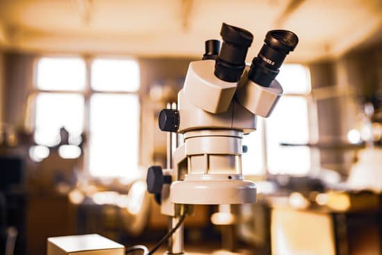
What year did the microscope invented? Lens Crafters Circa 1590: Invention of the Microscope. Every major field of science has benefited from the use of some form of microscope, an invention that dates back to the late 16th century and a modest Dutch eyeglass maker named Zacharias Janssen.
When was the first microscope invented? It’s not clear who invented the first microscope, but the Dutch spectacle maker Zacharias Janssen (b. 1585) is credited with making one of the earliest compound microscopes (ones that used two lenses) around 1600.
When was the first microscope discovered and by whom? 1590: Two Dutch spectacle-makers and father-and-son team, Hans and Zacharias Janssen, create the first microscope.
Who invented the microscope in 1932? 1900s. 1903: Richard Zsigmondy developed the ultramicroscope capable of studying objects below the wavelength of light. For this, he won the Nobel Prize in Chemistry in 1925. 1932: Frits Zernike invented the phase-contrast microscope that allowed for the study of colorless and transparent biological materials.
What year did the microscope invented? – Related Questions
Who invented the microscope lens?
Grinding glass to use for spectacles and magnifying glasses was commonplace during the 13th century. In the late 16th century several Dutch lens makers designed devices that magnified objects, but in 1609 Galileo Galilei perfected the first device known as a microscope.
How to count platelets under microscope?
An estimated platelet count can be done quickly with a blood smear evaluated at 100× magnification. At this magnification, the platelet count/µL of blood can be approximated by multiplying the number of platelets seen in one microscope field by 15,000.
Which microscopes use uv light?
UV microscopes have commonly been used in fluorescent microscopy. In this case, the UV light that reflects the image of the sample stains to the fluorescence to create an image that can be viewed.
Is a immersion oil microscope better?
Microscope immersion oil is used in light microscopy to improve imaging. The use of microscope immersion oil as part of a microscope lens system will produce a brighter and sharper image than a similar design not using immersion oil.
How did the microscope influence forensics?
The microscope has greatly affected the field of forensic science. Forensics is a field of science used to gather and analyze evidence to establish facts that are used in a legal scenario. The microscope is used to examine evidence collected in a crime scene that may have information not visible to the human eye.
How does letter e look under the microscope?
The letter “e” appears upside down and backwards under a microscope. Either, diatoms are single celled, or they do not have a cell wall.
How to increase light on a microscope?
The microscope rheostat control can be found on the side of the compound microscope body. It will typically be a knob that is turned clockwise in order to increase the light intensity, or counter-clockwise to reduce the light.
Does orientation change under a microscope?
The optics of a microscope’s lenses change the orientation of the image that the user sees. A specimen that is right-side up and facing right on the microscope slide will appear upside-down and facing left when viewed through a microscope, and vice versa.
What can you see with 40x microscope?
At 40x magnification you will be able to see 5mm. At 100x magnification you will be able to see 2mm. At 400x magnification you will be able to see 0.45mm, or 450 microns. At 1000x magnification you will be able to see 0.180mm, or 180 microns.
Who found compound microscope?
A Dutch father-son team named Hans and Zacharias Janssen invented the first so-called compound microscope in the late 16th century when they discovered that, if they put a lens at the top and bottom of a tube and looked through it, objects on the other end became magnified.
Are nerve endings microscopic?
Nerve endings in skin are involved in physiological processes such as sensing1 as well as in pathological processes such as neuropathic pain2. Their close-to-surface positioning facilitates microscopic imaging of skin nerve endings in living intact animal.
What are organisms you can only see with a microscope?
Bacteria are the smallest micro-organisms, ranging from between 0.0001 mm and 0.001 mm in size. Phytoplankton and protozoa range from about 0.001 mm to about 0.25 mm. The largest phytoplankton and protozoa can be seen with the naked eye, but most can only been seen under a microscope.
Who made the very first rudimentary microscope?
Lens Crafters Circa 1590: Invention of the Microscope. Every major field of science has benefited from the use of some form of microscope, an invention that dates back to the late 16th century and a modest Dutch eyeglass maker named Zacharias Janssen.
What is the highest magnification of a microscope?
Using the mathematical equations given above and the values for maximum numerical aperture attainable with the lenses of a light microscope it can be shown that the maximum useful magnification on a light microscope is between 1000X and 1500X. Higher magnification is possible, but resolution will not improve.
Did robert hooke invented the light microscope?
Although Hooke did not make his own microscopes, he was heavily involved with the overall design and optical characteristics. The microscopes were actually made by London instrument maker Christopher Cock, who enjoyed a great deal of success due to the popularity of this microscope design and Hooke’s book.
What two parts of the microscope adjust lighting?
Most high quality microscopes include an Abbe condenser with an iris diaphragm. Combined, they control both the focus and quantity of light applied to the specimen. Condenser Focus Knob moves the condenser up or down to control the lighting focus on the specimen.
When to use a compound microscope?
Typically, a compound microscope is used for viewing samples at high magnification (40 – 1000x), which is achieved by the combined effect of two sets of lenses: the ocular lens (in the eyepiece) and the objective lenses (close to the sample).
What kind of microscope is robert hookes?
He designed his own light microscope, which used multiple glass lenses to light and magnify specimens. Under his microscope, Hooke examined a diverse collection of organisms.
Can a phone be a microscope?
You may not think of your mobile phone as being anything like a microscope, but it has almost all the parts you need. The lens and camera sensor are arranged exactly as they would be inside a microscope – all you need to do to get some magnification is stick another lens in front.
What does the prism do on a microscope?
In modern microscopes equipped with binocular eyepiece tubes, prisms are also utilized to change the line of sight direction from vertical to a more convenient 45-degree angle.
How to improve resolving power of a microscope?
One way of increasing the optical resolving power of the microscope is to use immersion liquids between the front lens of the objective and the cover slip. Most objectives in the magnification range between 60x and 100x (and higher) are designed for use with immersion oil.

