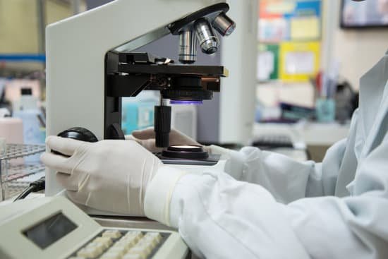
What’s a unicellular organism under a microscope? Unicellular organisms are made up of only one cell that carries out all of the functions needed by the organism, while multicellular organisms use many different cells to function. Unicellular organisms include bacteria, protists, and yeast.
Can you see a unicellular organism with a microscope? Unicellular organisms, or organisms that are made of only one cell. They are so small that they usually cannot be seen without a microscope. A unicellular organism behaves differently than a cell in a multicellular organism.
What are microscopic unicellular organisms? A unicellular organism is an organism that consists of a single cell. … Amoebas, bacteria, and plankton are just some types of unicellular organisms. They are typically microscopic and cannot be seen with the naked eye.
What are 5 examples of unicellular organisms? Unicellular organisms are composed of a single cell, unlike multicellular organisms that are made of many cells. This means that they each live and carry out all of their life processes as one single cell. Most unicellular organisms are microscopic; however, some are visible to the naked eye.
What’s a unicellular organism under a microscope? – Related Questions
What are the structure of dentin under the microscope?
Structure. Unlike enamel, dentin may be demineralized and stained for histological study. Dentin consists of microscopic channels, called dentinal tubules, which radiate outward through the dentin from the pulp to the exterior cementum or enamel border.
Is microscopic polyangiitis hereditary?
What causes microscopic polyangiitis (MPA)? The cause of MPA is unknown. It is not contagious, does not usually run in families, and is not a form of cancer .
How powerful of a microscope to see atoms?
An electron microscope can be used to magnify things over 500,000 times, enough to see lots of details inside cells. There are several types of electron microscope. A transmission electron microscope can be used to see nanoparticles and atoms.
What microscope is used to see white blood cells?
Thin sections of white blood cells from human being, guinea pigs and cats are examined under the electron microscope.
How does a microscope achieve magnification?
In simple magnification, light from an object passes through a biconvex lens and is bent (refracted) towards your eye. … Both of these contribute to the magnification of the object. The eyepiece lens usually magnifies 10x, and a typical objective lens magnifies 40x.
What do you call someone who works with microscopes?
Some types of biologists frequently use microscopes in research. For example, microbiologists use microscopes to study organisms that are too small to be seen with the naked eye, such as bacteria.
What are the magnifications on a light microscope?
Light microscopes combine the magnification of the eyepiece and an objective lens. Calculate the magnification by multiplying the eyepiece magnification (usually 10x) by the objective magnification (usually 4x, 10x or 40x). The maximum useful magnification of a light microscope is 1,500x.
Are bacteria microscopic organisms?
Technically a microorganism or microbe is an organism that is microscopic. The study of microorganisms is called microbiology. Microorganisms can be bacteria, fungi, archaea or protists. The term microorganisms does not include viruses and prions, which are generally classified as non-living.
Do u need a microscope to gram stain analysis?
Your sample will be placed on a slide and treated with the Gram stain. A laboratory professional will examine the slide under a microscope. … If the bacteria was colored pink or red, it means you likely have a Gram-negative infection.
How does a microscope magnify an image quizlet?
A compound light microscope uses two lenses at the same time to view objects-the objective lens, which gathers light and magnifies the image of the object, and the ocular lens, which one looks through and which further magnifies the image.
What do chromosomes look like under the microscope?
During most of the cell cycle, interphase, the chromosomes are somewhat less condensed and are not visible as individual objects under the light microscope. However during cell division, mitosis, the chromosomes become highly condensed and are then visible as dark distinct bodies within the nuclei of cells.
What type of microscopic algae is responsible for red tides?
At least three species of dinoflagellates and one diatom species are responsible for the toxic mess of red tides in the United States. These microscopic forms of algae produce toxins that can sicken humans and be fatal for marine animals.
What are the different types of bulb in microscope?
There are traditional projection bulbs, festoon bulbs,fluorescent ring bulbs, halogen and tungsten bulbs, and even mercury vapor bulbs. Some microscope bulbs aren’t bulbs at all but instead use LED clusters to illuminate samples for observation.
Why are images reversed in a microscope?
The eyepiece of the microscope contains a 10x magnifying lens, so the 10x objective lens actually magnifies 100 times and the 40x objective lens magnifies 400 times. There are also mirrors in the microscope, which cause images to appear upside down and backwards.
What is an electron microscope ks3?
An electron microscope is a scientific instrument which uses a beam of electrons to examine objects on a very fine scale. In an optical microscope, the wavelength of light limits the maximum magnification that is possible.
Can a student microscope see bacteria?
The answer is a careful “yes, but”. Generally speaking, it is theoretically and practically possible to see living and unstained bacteria with compound light microscopes, including those microscopes which are used for educational purposes in schools.
What describes the features of an electron microscope?
The electron microscope uses a beam of electrons and their wave-like characteristics to magnify an object’s image, unlike the optical microscope that uses visible light to magnify images. … This stream is confined and focused using metal apertures and magnetic lenses into a thin, focused, monochromatic beam.
What is used for calibration of a compound microscope?
The stage micrometer is used to calibrate an eyepiece reticle when making measurements with a microscope. Eyepiece Reticle (or reticule) -a small piece of glass with a ruler etched into it that fits into a microscope eyepiece. This ruler is used to make measurements of objects viewed through the microscope.
What controls the light on a microscope?
Iris Diaphragm controls the amount of light reaching the specimen. It is located above the condenser and below the stage. Most high quality microscopes include an Abbe condenser with an iris diaphragm. Combined, they control both the focus and quantity of light applied to the specimen.
What can be observed using the light microscope?
Light microscopes can be adapted to examine specimens of any size, whole or sectioned, living or dead, wet or dry, hot or cold, and static or fast-moving. They offer a wide range of contrast techniques, providing information on the physical, chemical, and biological attributes of specimens.
Can you see giardia under microscope?
In bright-field microscopy, cysts appear ovoid to ellipsoid in shape and usually measure 11 to 14 µm (range: 8 to 19 µm). Immature and mature cysts have 2 and 4 nuclei, respectively. Intracytoplasmic fi brils are visible in cysts.

