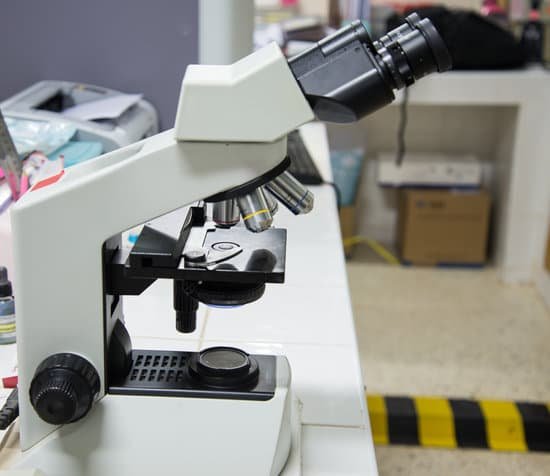
When was the first microscope invented and by whom? Lens Crafters Circa 1590: Invention of the Microscope. Every major field of science has benefited from the use of some form of microscope, an invention that dates back to the late 16th century and a modest Dutch eyeglass maker named Zacharias Janssen.
When was the first microscope made and by whom? It’s not clear who invented the first microscope, but the Dutch spectacle maker Zacharias Janssen (b. 1585) is credited with making one of the earliest compound microscopes (ones that used two lenses) around 1600.
Who invented microscope at first? A Dutch father-son team named Hans and Zacharias Janssen invented the first so-called compound microscope in the late 16th century when they discovered that, if they put a lens at the top and bottom of a tube and looked through it, objects on the other end became magnified.
Who invented the microscope in 1666? Antoni Van Leeuwenhoek (1635-1723) was a Dutch tradesman who became interested in microscopy while on a visit to London in 1666. Returning home, he began making simple microscopes of the sort that Robert Hooke had described in his, Micrographia, and using them to discover objects invisible to the naked eye.
When was the first microscope invented and by whom? – Related Questions
Why doctors use microscopes?
The cells that appeared under the microscope were epithelial cells, which line the surfaces of our organs and blood vessels. Doctors and clinicians still use medical microscopes to identify these types of cells, which can often tell us when something is going wrong in our bodies.
What year did robert hooke invent his microscope?
Robert Hooke’s Microscope. Robert Hook refined the design of the compound microscope around 1665 and published a book titled Micrographia which illustrated his findings using the instrument.
Why can’t mitochondria be seen with a light microscope?
However, most organelles are not clearly visible by light microscopy, and those that can be seen (such as the nucleus, mitochondria and Golgi) can’t be studied in detail because their size is close to the limit of resolution of the light microscope.
What does a iris diaphragm do in a microscope?
Iris Diaphragm controls the amount of light reaching the specimen. It is located above the condenser and below the stage. Most high quality microscopes include an Abbe condenser with an iris diaphragm. Combined, they control both the focus and quantity of light applied to the specimen.
When was the compound light microscope invented?
received credit for inventing the compound microscope about 1590. The first portrayal of a microscope was drawn about 1631 in the Netherlands.
What does urinalysis with reflex microscopic tell?
Urinalysis with Reflex to Microscopic – Dipstick urinalysis measures chemical constituents of urine. Microscopic examination helps to detect the presence of cells, bacteria, yeast and other formed elements.
How many spores per ascus are visible in the microscope?
In ascomycetes the spores are produced within microscopic cells called asci. The asci vary in shape from cylindric to spherical. Commonly, each ascus holds eight spores – but there are species with just one spore per ascus and others with over a hundred spores per ascus.
How is a light microscope work used?
The light microscope is an instrument for visualizing fine detail of an object. It does this by creating a magnified image through the use of a series of glass lenses, which first focus a beam of light onto or through an object, and convex objective lenses to enlarge the image formed.
What is an inverted optical microscope?
An inverted microscope is a microscope with its light source and condenser on the top, above the stage pointing down, while the objectives and turret are below the stage pointing up. It was invented in 1850 by J. Lawrence Smith, a faculty member of Tulane University (then named the Medical College of Louisiana).
How to avoid bubbles in permanent microscope slides?
Place a sample on the slide. Using a pipette, place a drop of water on the specimen. Then place on edge of the cover slip over the sample and carefully lower the cover slip into place using a toothpick or equivalent. This method will help prevent air bubbles from being trapped under the cover slip.
Is light microscope and compound microscope the same?
The common light microscope used in the laboratory is called a compound microscope because it contains two types of lenses that function to magnify an object. The lens closest to the eye is called the ocular, while the lens closest to the object is called the objective.
How to clean meiji microscope?
Using the cotton end of the stick, start at the center of the lens using a circular motion and work your way to the outer edge. Gently wipe off any excess liquid with another dry lint-free swab. Using another lint-free swab, gently wipe off any residue on the glass.
What does it mean to put someone under a microscope?
phrase. DEFINITIONS1. if someone or something is under the microscope, people are examining them very carefully. The whole legal system should be put under the microscope. Synonyms and related words.
What can transmission electron microscopes do?
Transmission electron microscopes (TEM) are microscopes that use a particle beam of electrons to visualize specimens and generate a highly-magnified image. TEMs can magnify objects up to 2 million times. In order to get a better idea of just how small that is, think of how small a cell is.
What are the objectives of a compound microscope?
Standard objectives include 4x, 10x, 40x and 100x although different power objectives are available. Coarse and Fine Focus knobs are used to focus the microscope. Increasingly, they are coaxial knobs – that is to say they are built on the same axis with the fine focus knob on the outside.
Which is a microscopic examination of living tissue?
Biopsy – The removal and examination, usually microscopic, of tissue from the living body, performed to establish precise diagnosis.
What microscope can see dust mites?
As I mentioned earlier, dust mites are microscopic creatures which cannot be seen by a naked human eye. However, they can easily be seen under a microscope with at least a 10x magnification lens. Most standard microscopes have 10x magnification eyepieces.
Can’t see anything inside my microscope?
If you cannot see anything, move the slide slightly while viewing and focusing. If nothing appears, reduce the light and repeat step 4. Once in focus on low power, center the object of interest by moving the slide. Rotate the objective to the medium power and adjust the fine focus only.
What were electron microscopes originally used for?
The ultimate goal was atomic resolution – the ability to see atoms – but this would have to be approached incrementally over the course of decades. The earliest microscopes merely proved the concept: electron beams could, indeed, be tamed to provide visible images of matter.
Why do microscopes alter images?
Microscopes invert images which makes the picture appear to be upside down. The reason this happens is that microscopes use two lenses to help magnify the image. Some microscopes have additional magnification settings which will turn the image right-side-up.
Where are the objective lenses located on a compound microscope?
The lenses that are attached to the nosepiece of a compound light microscope are referred to as the objective lens. A compound light microscope will have several (usually four) pieces of objective lenses with different magnification powers. The common magnification powers are 4x, 10x, 40x, and 100x.

