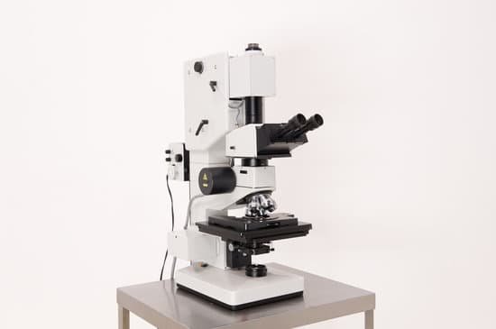
Where do you place a slide on a microscope? Place the microscope slide on the stage (6) and fasten it with the stage clips. Look at the objective lens (3) and the stage from the side and turn the focus knob (4) so the stage moves upward. Move it up as far as it will go without letting the objective touch the coverslip.
Which part of the microscope holds the specimen? The stage is a platform for the slides, which hold the specimen. The stage typically has a stage clip on either side to hold the slide firmly in place. Some microscopes have a mechanical stage, with adjustment knobs that allow for more precise positioning of slides.
What is used to hold the microscope? The three basic, structural components of a compound microscope are the head, base and arm. Arm connects to the base and supports the microscope head. It is also used to carry the microscope.
What do you use to hold a specimen to view under the microscope? A microscope slide is a thin flat piece of glass, typically 75 by 26 mm (3 by 1 inches) and about 1 mm thick, used to hold objects for examination under a microscope. Typically the object is mounted (secured) on the slide, and then both are inserted together in the microscope for viewing.
Where do you place a slide on a microscope? – Related Questions
Can a microscope be used to see atoms?
An electron microscope can be used to magnify things over 500,000 times, enough to see lots of details inside cells. There are several types of electron microscope. A transmission electron microscope can be used to see nanoparticles and atoms.
How to preserve microscope specimens?
To keep your prepared microscope slides in good condition, always store them in a container made for the purpose and away from heat and bright light. The ideal storage area is a cool, dark location, such as a closed cabinet in a temperature-controlled room. Stained slides naturally fade over time.
What does x10 mean on a microscope?
100X (this means that the image being viewed will appear to be 100 times its actual size).
How long was the first microscope?
1619 — Earliest recorded description of a compound microscope, Dutch Ambassador Willem Boreel sees one in London in the possession of Dutch inventor Cornelis Drebbel, an instrument about eighteen inches long, two inches in diameter, and supported on 3 brass dolphins.
Which microscope has magnification up to 1 million x?
An SEM can magnify a sample by about one million times (1,000,000x) at the most. Because a sample can be used in its natural state, the SEM is the easiest electron microscope to use.
How does sperm look in microscope?
The air-fixed, stained spermatozoa are observed under a bright-light microscope at 400x or 1000x magnification. Their viability and mor- phology can be analysed at the same time. Those appearing red-pink in colour have a damaged membrane whereas white sperm are viable, as in Photo 2.
What is the most magnified microscope?
Lawrence Berkeley National Labs just turned on a $27 million electron microscope. Its ability to make images to a resolution of half the width of a hydrogen atom makes it the most powerful microscope in the world.
What is meant by compound microscope?
A compound microscope is a microscope that uses multiple lenses to enlarge the image of a sample. … Compound microscopes usually include exchangeable objective lenses with different magnifications (e.g 4x, 10x, 40x and 60x), mounted on a turret, to adjust the magnification.
How good a microscope do you use to see bacteria?
Bacteria are too small to see without the aid of a microscope. While some eucaryotes, such as protozoa, algae and yeast, can be seen at magnifications of 200X-400X, most bacteria can only be seen with 1000X magnification. This requires a 100X oil immersion objective and 10X eyepieces..
What are the four lenses of the microscope?
A typical compound microscope will have four objective lenses: one scanning lens, low-power lens, high-power lens, and an oil-immersion lens.
How to calculate magnification on microscope?
To figure the total magnification of an image that you are viewing through the microscope is really quite simple. To get the total magnification take the power of the objective (4X, 10X, 40x) and multiply by the power of the eyepiece, usually 10X.
What is the microscope nosepiece?
Revolving Nosepiece or Turret: This is the part that holds two or more objective lenses and can be rotated to easily change power. Objective Lenses: Usually you will find 3 or 4 objective lenses on a microscope. They almost always consist of 4X, 10X, 40X and 100X powers.
How to carry and store a microscope?
Always keep your microscope covered when not in use. Always carry a microscope with both hands. Grasp the arm with one hand and place the other hand under the base for support.
What type of microscope would you use to observe cilia?
TEM passes an electron beam through a thin, fixed sample and has been a valuable tool for studying ciliated respiratory epithelium because of its ability to achieve subnanometer resolution (32, 100, 146).
Do microscopes use lenses?
While the modern microscope has many parts, the most important pieces are its lenses. It is through the microscope’s lenses that the image of an object can be magnified and observed in detail. … When light reflects off of an object being viewed under the microscope and passes through the lens, it bends towards the eye.
How many types of electron microscope?
There are two main types of electron microscope – the transmission EM (TEM) and the scanning EM (SEM). The transmission electron microscope is used to view thin specimens (tissue sections, molecules, etc) through which electrons can pass generating a projection image.
Is there anything smaller than microscopic?
In short, all objects that are too small for you to see are microscopic, but there is no precise line between microscopic and non-microscopic. For example, a grain of sand may technically not be microscopic, because some of them you can see with a naked eye.
What is the coarse adjustment knob on a microscope?
COARSE ADJUSTMENT KNOB — A rapid control which allows for quick focusing by moving the objective lens or stage up and down. It is used for initial focusing.
What is the microscopic functional unit of the kidney?
The nephron is the functional unit of the kidney. The glomerulus and convoluted tubules of the nephron are located in the cortex of the kidney, while the collecting ducts are located in the pyramids of the kidney’s medulla.
How to calculate field diameter in a microscope?
The field size or diameter at a given magnification is calculated as the field number divided by the objective magnification. If the ×40 objective is used, the diameter of the field of view becomes 20 mm/40 (compared with no objective) or 0.5 mm.

