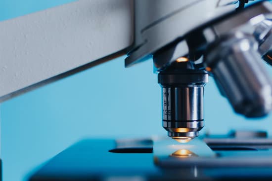
Which cell organelles can be viewed with light microscopes? Note: The nucleus, cytoplasm, cell membrane, chloroplasts and cell wall are organelles which can be seen under a light microscope.
What organisms can be seen with a light microscope? Explanation: You can see most bacteria and some organelles like mitochondria plus the human egg. You can not see the very smallest bacteria, viruses, macromolecules, ribosomes, proteins, and of course atoms.
Can light microscopes be used to observe organelles of a cell? Since most cells are between 1 and 100 μm in diameter, they can be observed by light microscopy, as can some of the larger subcellular organelles, such as nuclei, chloroplasts, and mitochondria.
What are the types of microscopy? So, we can think of the microscopic scale as being from a millimetre (10-3 m) to a ten-millionth of a millimetre (10-10 m). Even within the microscopic scale, there are immense variations in the size of objects.
Which cell organelles can be viewed with light microscopes? – Related Questions
What does the arm do on the microscope?
Arm connects to the base and supports the microscope head. It is also used to carry the microscope.
Is microscopic hematuria normal?
Conclusions. Asymptomatic microscopic hematuria in women is common; however, it is less likely to be associated with urinary tract malignancy among women than men. For women, being older than 60 years, having a history of smoking, and having gross hematuria are the strongest predictors of urologic cancer.
How is a microscope used in science?
A microscope is an instrument that is used to magnify small objects. Some microscopes can even be used to observe an object at the cellular level, allowing scientists to see the shape of a cell, its nucleus, mitochondria, and other organelles.
What is the purpose of the condenser on a microscope?
On upright microscopes, the condenser is located beneath the stage and serves to gather wavefronts from the microscope light source and concentrate them into a cone of light that illuminates the specimen with uniform intensity over the entire viewfield.
Who invented the compact microscope?
Every major field of science has benefited from the use of some form of microscope, an invention that dates back to the late 16th century and a modest Dutch eyeglass maker named Zacharias Janssen.
Did robert hooke invent microscopes?
Although Hooke did not make his own microscopes, he was heavily involved with the overall design and optical characteristics. The microscopes were actually made by London instrument maker Christopher Cock, who enjoyed a great deal of success due to the popularity of this microscope design and Hooke’s book.
What is refractive index in microscope?
Refractive Index (Index of Refraction) is a value calculated from the ratio of the speed of light in a vacuum to that in a second medium of greater density. The refractive index variable is most commonly symbolized by the letter n or n’ in descriptive text and mathematical equations.
What is the eyepiece of a microscope called quizlet?
cover slip. … The total magnifying power of a microscope is the product of the magnifying power of the lens in the eyepiece (the ocular) and the magnifying power of the lens in the objective.
What is the angular magnification of the microscope?
The angular magnification of a compound microscope is the ratio of the angle subtended by the final image at the eye to the angle subtended by the object at the eye, when both are placed at the least distance of distinct vision. This is the required expression for angular magnification.
What are the limitations of microscopic firearms analysis?
The biggest limitation would be the condition of the evidence. If the evidence (bullets and cartridge cases) is too damaged or mutilated to reveal sufficient individual characteristics, then no comparison can be made. The lack of a suspected firearm also presents limitations for the examiner’s conclusions.
What does the base of a microscope support?
Base: The bottom of the microscope, used for support. Illuminator: A steady light source (110 volts) used in place of a mirror. If your microscope has a mirror, it is used to reflect light from an external light source up through the bottom of the stage.
What does the term working distance mean microscope?
■ Working Distance (W.D.) The distance between the front end of a microscope objective and the. surface of the workpiece at which the sharpest focusing is obtained.
What does it mean when you have microscopic hematuria?
“Microscopic” means something is so small that it can only be seen through a special tool called a microscope. “Hematuria” means blood in the urine. So, if you have microscopic hematuria, you have red blood cells in your urine. These blood cells are so small, though, you can’t see the blood when you urinate.
Why should you start from low power lens in microscope?
When using a light microscope it’s important to start with the low power objective lens as the field of view will be wider, increasing the number of cells you are able to see. This makes it easier to find what you’re looking for.
How much does a confocal microscope cost?
Prices may range anywhere from under $750 to over $89,000, depending on features. When considering confocal microscopes, there are more distinctive features associated with different models.
Which microscope objective has the smallest working distance?
The oil immersion lens has the smallest working distance and one runs the risk of striking the slide with the lens when trying to achieve focus.
How do you increase the resolution on your microscope?
To achieve the maximum (theoretical) resolution in a microscope system, each of the optical components should be of the highest NA available (taking into consideration the angular aperture). In addition, using a shorter wavelength of light to view the specimen will increase the resolution.
Is hyphae microscopic?
Hyphae, as mentioned, grow from the spore/germ. … While some of these tubular structures can be seen with the naked eye (in large numbers) an individual hypha is a microscopic tube like structures that contain a cytoplasm (multinucleate cytoplasm) that is surrounded by a plasma membrane.
When was a electron microscope first used?
It was Ernst Ruska and Max Knoll, a physicist and an electrical engineer, respectively, from the University of Berlin, who created the first electron microscope in 1931. This prototype was able to produce a magnification of four-hundred-power and was the first device to show what was possible with electron microscopy.
What is the total magnification of a specimen in microscope?
Total Magnification: To figure the total magnification of an image that you are viewing through the microscope is really quite simple. To get the total magnification take the power of the objective (4X, 10X, 40x) and multiply by the power of the eyepiece, usually 10X.

