Which is smaller than microscopic or the submicroscopic? As adjectives the difference between submicroscopic and macroscopic. is that submicroscopic is smaller than microscopic; too small to be seen even with a microscope while macroscopic is visible to the unassisted eye.
What is smaller submicroscopic or microscopic? As adjectives the difference between microscopic and submicroscopic. is that microscopic is of, or relating to microscopes or microscopy; microscopal while submicroscopic is smaller than microscopic; too small to be seen even with a microscope.
Is there anything smaller than microscopic? In short, all objects that are too small for you to see are microscopic, but there is no precise line between microscopic and non-microscopic. For example, a grain of sand may technically not be microscopic, because some of them you can see with a naked eye.
Is microscopic or nanoscopic smaller? As adjectives the difference between microscopic and nanoscopic. is that microscopic is of, or relating to microscopes or microscopy; microscopal while nanoscopic is having a scale expressed in nanometers.
Which is smaller than microscopic or the submicroscopic? – Related Questions
What is scanning electron microscope ppt?
SCANNING ELECTRON MICROSCOPE (SEM) A scanning electron microscope (SEM) is a type of electron microscope that images a sample by scanning it with a high-energy beam of electrons in a raster scan pattern.
How does dna look under a microscope?
Given that DNA molecules are found inside the cells, they are too small to be seen with the naked eye. … While it is possible to see the nucleus (containing DNA) using a light microscope, DNA strands/threads can only be viewed using microscopes that allow for higher resolution.
Did hooke invent the microscope?
Although Hooke did not make his own microscopes, he was heavily involved with the overall design and optical characteristics. The microscopes were actually made by London instrument maker Christopher Cock, who enjoyed a great deal of success due to the popularity of this microscope design and Hooke’s book.
Which muscle types appear striated when viewed under a microscope?
Skeletal muscles are long and cylindrical in appearance; when viewed under a microscope, skeletal muscle tissue has a striped or striated appearance. The striations are caused by the regular arrangement of contractile proteins (actin and myosin).
What does microscopic blood in your urine mean?
Microscopic urinary bleeding is a common symptom of glomerulonephritis, an inflammation of the kidneys’ filtering system. Glomerulonephritis may be part of a systemic disease, such as diabetes, or it can occur on its own.
How to calculate field of view light microscope?
For instance, if your eyepiece reads 10X/22, and the magnification of your objective lens is 40. First, multiply 10 and 40 to get 400. Then divide 22 by 400 to get a FOV diameter of 0.055 millimeters.
What does human hair look like under a microscope?
Human hair under a microscope resembles animal fur. It looks like a tube filled with keratin (pigment) and covered with small scales outside. If these scales are growing tightly, hair looks smooth and shiny. Dull and unruly hair looks different under a microscope – the scales are disheveled and tumbled.
What is the function of the lamp on a microscope?
lamp – produces the light (Typically, lamps are tungsten-filament light bulbs. For specialized applications, mercury or xenon lamps may be used to produce ultraviolet light. Some microscopes even use lasers to scan the specimen.)
Which type of microscope provides the greatest resolution biology quizlet?
The electron microscope has greater resolution than the light microscope. A. Both the electron microscope and the light microscope use the same wavelengths for illumination. The light microscope uses light, and the electron microscope uses electrons.
Can atoms be seen under microscope?
Atoms are really small. So small, in fact, that it’s impossible to see one with the naked eye, even with the most powerful of microscopes. … Now, a photograph shows a single atom floating in an electric field, and it’s large enough to see without any kind of microscope.
Why are there multiple objectives on a microscope?
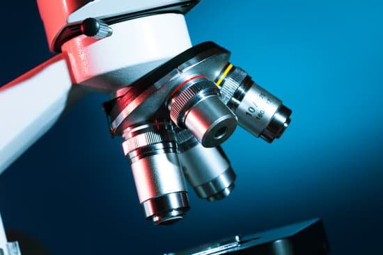
Although a microscope objective is sometimes called the objective lens, it usually contains multiple lenses. The higher the numerical aperture and the higher the required image quality, the more sophisticated designs are needed.
What kind of steroids do they use for microscopic colitis?
Steroids work by decreasing inflammation and reducing the activity of the immune system. The two steroids most often prescribed for microscopic colitis are budesonide (Entocort®) and prednisone. Budesonide is believed to be the safest and most effective medication for treating microscopic colitis.
What do you adjust first on a microscope?
FIRST CLOSE THE FIELD DIAPHRAGM TO IT’S SMALLEST OPENING BY TURNING THE KNURLED ADJUSTMENT KNOB LOCATED BEHIND THE FIELD LENS ASSEMBLY IN THE BASE OF YOUR MICROSCOPE.
How does a sem microscope work?
The SEM is an instrument that produces a largely magnified image by using electrons instead of light to form an image. A beam of electrons is produced at the top of the microscope by an electron gun. … Once the beam hits the sample, electrons and X-rays are ejected from the sample.
How to look in a microscope?
Look at the objective lens (3) and the stage from the side and turn the focus knob (4) so the stage moves upward. Move it up as far as it will go without letting the objective touch the coverslip. Look through the eyepiece (1) and move the focus knob until the image comes into focus.
How does an object look under a microscope?
A simple light microscope manipulates how light enters the eye using a convex lens, where both sides of the lens are curved outwards. When light reflects off of an object being viewed under the microscope and passes through the lens, it bends towards the eye. This makes the object look bigger than it actually is.
What is microscope and its uses?
A microscope is an instrument that can be used to observe small objects, even cells. The image of an object is magnified through at least one lens in the microscope. This lens bends light toward the eye and makes an object appear larger than it actually is.
What is the scanning objective on a microscope?
A scanning objective lens provides the lowest magnification power of all objective lenses. … The name “scanning” objective lens comes from the fact that they provide observers with about enough magnification for a good overview of the slide, essentially a “scan” of the slide.
How are microscopes used in clinical settings?
Microscopes are typically used in surgical fields such as dentistry, plastic surgery, ophthalmic surgery which involves the eyes, ear, nose and throat (ENT) surgery, and neurosurgery. Without microscopes, several diseases and illnesses can’t be identified, particularly cellular diseases.
Why do vets use microscopes?
Microscopes have many uses in veterinary medicine. When we do a fecal exam, we use them to look for eggs of intestinal parasites. They are helpful for diagnosing ear problems. By looking at a slide, we can check for ear mites, bacterial infections, and yeast infections.

