Which microscope is best for viewing protein? Electron microscope can be used to see protein molecules. An electron microscope that generates high-energy electrons gives an electronic image to observe protein molecules. The high-resolution pictures ensure the arrangement and structure of biomolecules.
Can you see proteins under a microscope? Using superresolution fluorescence microscopy, one can pinpoint the location of proteins at a resolution of 20 nm or even less.
Can Tem See proteins? Metal-tagging TEM using MT shows proteins in cells at molecular scale resolution. METTEM exceeds sensitivity of immunogold detection by orders of magnitude. METTEM revealed virus-induced cell structures and organelles not seen before.
Why do we need standard precautions? Universal precautions (UP), originally recommended by the CDC in the 1980s, was introduced as an approach to infection control to protect workers from HIV, HBV, and other bloodborne pathogens in human blood and certain other body fluids, regardless of a patients’ infection status.
Which microscope is best for viewing protein? – Related Questions
Who made the stereo microscope?
The first stereoscopic-style microscope having twin eyepieces and matching objectives was designed and built by Cherubin d’Orleans in 1671, but the instrument was actually a pseudostereoscopic system that achieved image erection only by the application of supplemental lenses.
What is maximum resolution in micrometers of a light microscope?
The diffraction limits the resolution to approximately 0.2 µm. … The resolution of the light microscope cannot be small than the half of the wavelength of the visible light, which is 0.4-0.7 µm. When we can see green light (0.5 µm), the objects which are, at most, about 0.2 µm.
What is the function of the nosepiece on a microscope?
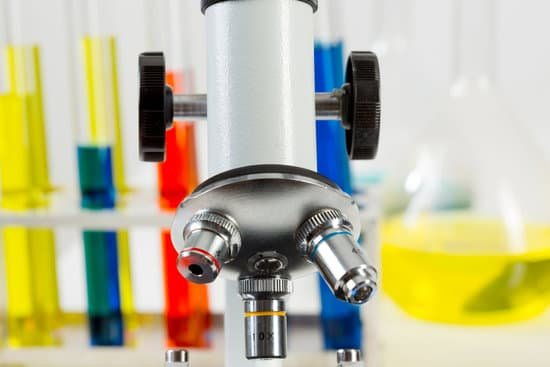
Revolving Nosepiece or Turret: This is the part that holds two or more objective lenses and can be rotated to easily change power. Objective Lenses: Usually you will find 3 or 4 objective lenses on a microscope. They almost always consist of 4X, 10X, 40X and 100X powers.
What increases or decreases the light intensity on a microscope?
There are essentially three ways to vary the brightness; by increasing or decreasing the light intensity (using the on/off knob), by moving the condenser lens closer to or farther from the object using the condenser adjustment knob, and/or by opening/closing the iris diaphragm.
How a stereo microscope works?
A stereo model is an optical microscope that functions at a low magnification. It works by using two separate optical paths instead of just one. … The lighting is also different than on other types of microscopes. It uses reflected, or episcopic, illumination to light up specimens.
How to work out the total magnification of a microscope?
To figure the total magnification of an image that you are viewing through the microscope is really quite simple. To get the total magnification take the power of the objective (4X, 10X, 40x) and multiply by the power of the eyepiece, usually 10X. Who uses microscopes?
Which microscope doesn’t show the image upside down?
Quite a few microscopes, including electron microscopes and digital microscopes, will not show you inverted images. Binocular and dissecting microscopes will also not show an inverted image because of their increased level of magnification.
What cell organelles cannot be seen with a light microscope?
Some cell parts, including ribosomes, the endoplasmic reticulum, lysosomes, centrioles, and Golgi bodies, cannot be seen with light microscopes because these microscopes cannot achieve a magnification high enough to see these relatively tiny organelles.
What is a monocular microscope used for?
Monocular microscopes, microscopes that are equipped with one eye piece, can magnify samples up to 1,000 times. If you need a microscope that magnifies at higher levels, a binocular microscope is right for you. Monocular microscopes are often used in classrooms and laboratories for observing slide samples.
What is an advantage of a light microscope?
Advantages. Inexpensive to buy and operate. Relatively small. Both living and dead specimens can be viewed. Little expertise is required in order to set up and use the microscope.
Which microscope can see inside of a cell?
Electrons have much a shorter wavelength than visible light, and this allows electron microscopes to produce higher-resolution images than standard light microscopes. Electron microscopes can be used to examine not just whole cells, but also the subcellular structures and compartments within them.
How to carry out microscopic malaria test?
Malaria parasites can be identified by examining under the microscope a drop of the patient’s blood, spread out as a “blood smear” on a microscope slide. Prior to examination, the specimen is stained (most often with the Giemsa stain) to give the parasites a distinctive appearance.
What is a microscope in science definition?
A microscope is an instrument that can be used to observe small objects, even cells. The image of an object is magnified through at least one lens in the microscope. This lens bends light toward the eye and makes an object appear larger than it actually is. 5 – 12+ Biology, Engineering.
How to view sperm on a microscope?
You can view sperm at 400x magnification. You do NOT want a microscope that advertises anything above 1000x, it is just empty magnification and is unnecessary. In order to examine semen with the microscope you will need depression slides, cover slips, and a biological microscope.
What a light microscope is used for?
Principles. The light microscope is an instrument for visualizing fine detail of an object. It does this by creating a magnified image through the use of a series of glass lenses, which first focus a beam of light onto or through an object, and convex objective lenses to enlarge the image formed.
How to improve resolution on a microscope?
To achieve the maximum (theoretical) resolution in a microscope system, each of the optical components should be of the highest NA available (taking into consideration the angular aperture). In addition, using a shorter wavelength of light to view the specimen will increase the resolution.
What size can a light microscope see?
Light microscopes let us look at objects as long as a millimetre (10-3 m) and as small as 0.2 micrometres (0.2 thousands of a millimetre or 2 x 10⁻⁷ m), whereas the most powerful electron microscopes allow us to see objects as small as an atom (about one ten-millionth of a millimetre or 1 angstrom or 10-10 m).
Do most cells under a microscope have visible chromosomes?
Chromosomes are not visible in the cell’s nucleus—not even under a microscope—when the cell is not dividing. However, the DNA that makes up chromosomes becomes more tightly packed during cell division and is then visible under a microscope.
When were microscopes used to study cells?
Indeed, the very discovery of cells arose from the development of the microscope: Robert Hooke first coined the term “cell” following his observations of a piece of cork with a simple light microscope in 1665 (Figure 1.23).
What type of microscope can see in angstroms?
The first electron microscope that can resolve features as small as half an Angstrom (0.05 nm) has been developed in the US.

