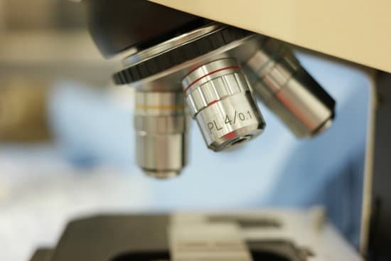
Which objective should be in place when storing the microscope? Always place the 4X objective over the stage and be sure the stage is at its lowest position before putting the microscope away. 9. Always turn off the light before putting the microscope away. 10.
Which objective should be in place when storing the microscope quizlet? The microscope should be put away with the scanning and objective lens in position.
Who invented microscope 1666? Antoni Van Leeuwenhoek (1635-1723) was a Dutch tradesman who became interested in microscopy while on a visit to London in 1666. Returning home, he began making simple microscopes of the sort that Robert Hooke had described in his, Micrographia, and using them to discover objects invisible to the naked eye.
Who invented microscope at first? Every major field of science has benefited from the use of some form of microscope, an invention that dates back to the late 16th century and a modest Dutch eyeglass maker named Zacharias Janssen.
Which objective should be in place when storing the microscope? – Related Questions
What microscope can see bacteria from water?
In order to actually see bacteria swimming, you’ll need a lens with at least a 400x magnification. A 1000x magnification can show bacteria in stunning detail.
What do you use to make microscope slides?
A wet mount requires a liquid, tweezers, pipette and paper towels. Although wet mounts can be used to prepare a significantly wide range of microscope slides, they provide a transitory window as the liquid will dehydrate and living specimens will die.
How to see sperm under microscope?
You can view sperm at 400x magnification. You do NOT want a microscope that advertises anything above 1000x, it is just empty magnification and is unnecessary. In order to examine semen with the microscope you will need depression slides, cover slips, and a biological microscope.
Which microscope ideal to look at bacteria?
In order to actually see bacteria swimming, you’ll need a lens with at least a 400x magnification. A 1000x magnification can show bacteria in stunning detail.
How does a compound microscope magnify an object?
A microscope is an instrument that can be used to observe small objects, even cells. The image of an object is magnified through at least one lens in the microscope. This lens bends light toward the eye and makes an object appear larger than it actually is.
What is microscopic lymphocytic colitis?
Lymphocytic colitis is one type of inflammatory bowel disease (IBD). IBD is a group of conditions that cause inflammation in either the small or large intestine. Lymphocytic colitis is a type of microscopic colitis. Microscopic colitis is inflammation of the large intestine that can only be seen through a microscope.
Is it dangerous to have microscopic blood in urine?
While in many instances the cause is harmless, blood in urine (hematuria) can indicate a serious disorder. Blood that you can see is called gross hematuria. Urinary blood that’s visible only under a microscope (microscopic hematuria) is found when your doctor tests your urine.
What is the slide holder on a microscope?
Description. The slide holder on a microscope is an important component for observing specimens by holding your slides secure and steady during observation.
What is used to calculate total magnification of a microscope?
To calculate total magnification, find the magnification of both the eyepiece and the objective lenses. The common ocular magnifies ten times, marked as 10x. The standard objective lenses magnify 4x, 10x and 40x. If the microscope has a fourth objective lens, the magnification will most likely be 100x.
What magnifies the image in a microscope by 10x?
100X (this means that the image being viewed will appear to be 100 times its actual size).
What is the iris diaphragm function on microscope?
Iris Diaphragm controls the amount of light reaching the specimen. It is located above the condenser and below the stage. Most high quality microscopes include an Abbe condenser with an iris diaphragm. Combined, they control both the focus and quantity of light applied to the specimen.
What is a objective lens used for a microscope?
The objective lens of a microscope is the one at the bottom near the sample. At its simplest, it is a very high-powered magnifying glass, with very short focal length. This is brought very close to the specimen being examined so that the light from the specimen comes to a focus inside the microscope tube.
What are the objectives used for on a microscope?
Objectives allow microscopes to provide magnified, real images and are, perhaps, the most complex component in a microscope system because of their multi-element design. Objectives are available with magnifications ranging from 2X – 200X.
Who made the microscope invented?
The development of the microscope allowed scientists to make new insights into the body and disease. It’s not clear who invented the first microscope, but the Dutch spectacle maker Zacharias Janssen (b. 1585) is credited with making one of the earliest compound microscopes (ones that used two lenses) around 1600.
What is stereo microscope used for?
A stereo microscope is used for low-magnification applications, allowing high-quality, 3D observation of subjects that are normally visible to the naked eye. In life science stereo microscope applications, this could involve the observation of insects or plant life.
How much does a sem microscope cost?
The price of electron microscopes can also vary by type of electron microscope. The cost of a scanning electron microscope (SEM) can range from $80,000 to $2,000,000. The cost of a transmission electron microscope (TEM) can range from $300,000 to $10,000,000.
Why are microscopes stored with the low power objective lens?
Objective lenses Always start and end your microscope session by placing the lowest power objective lens in position. This will make it easier to prevent crashing the objective lens into the slide. … Using the coarse focus with higher lenses may result in crashing the lens into the slide.
Do fluorescent microscopes have a high resolution?
Compared to other imaging techniques such as electron microscopy (EM), however, conventional fluorescence microscopy is limited by relatively low spatial resolution because of the diffraction of light.
Can you view an atom under a microscope?
Atoms are really small. So small, in fact, that it’s impossible to see one with the naked eye, even with the most powerful of microscopes. … Now, a photograph shows a single atom floating in an electric field, and it’s large enough to see without any kind of microscope.
Why was the microscope invented?
The invention of the microscope allowed scientists and scholars to study the microscopic creatures in the world around them. … Electron microscopes can provide pictures of the smallest particles but they cannot be used to study living things. Its magnification and resolution is unmatched by a light microscope.

