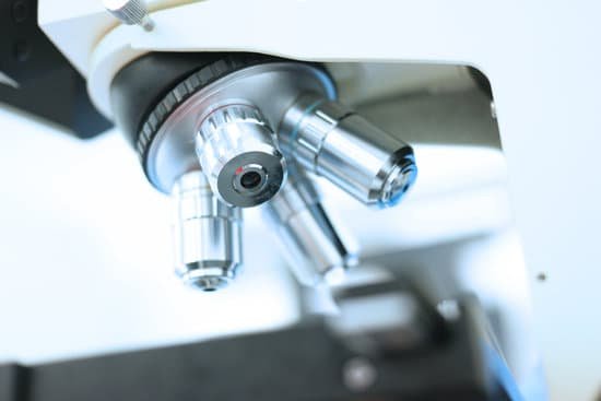
Which of these could you see with a light microscope? Explanation: You can see most bacteria and some organelles like mitochondria plus the human egg. You can not see the very smallest bacteria, viruses, macromolecules, ribosomes, proteins, and of course atoms.
What can you see with light microscope? Using a light microscope, one can view cell walls, vacuoles, cytoplasm, chloroplasts, nucleus and cell membrane. Light microscopes use lenses and light to magnify cell parts. However, they usually can achieve a maximum of 2000x magnification which is not sufficient to see many other tiny organelles.
What parts of the cell can be seen with a light microscope? Note: The nucleus, cytoplasm, cell membrane, chloroplasts and cell wall are organelles which can be seen under a light microscope.
What does a light microscope see an image? It is through the microscope’s lenses that the image of an object can be magnified and observed in detail. A simple light microscope manipulates how light enters the eye using a convex lens, where both sides of the lens are curved outwards.
Which of these could you see with a light microscope? – Related Questions
What does the condenser on your microscope do?
The substage condenser gathers light from the microscope light source and concentrates it into a cone of light that illuminates the specimen with uniform intensity over the entire viewfield.
Who was the first to observe microorganisms with a microscope?
The existence of microscopic organisms was discovered during the period 1665-83 by two Fellows of The Royal Society, Robert Hooke and Antoni van Leeuwenhoek. In Micrographia (1665), Hooke presented the first published depiction of a microganism, the microfungus Mucor.
How to determine microscope magnification?
To figure the total magnification of an image that you are viewing through the microscope is really quite simple. To get the total magnification take the power of the objective (4X, 10X, 40x) and multiply by the power of the eyepiece, usually 10X.
What is the most powerful microscope on the planet?
Lawrence Berkeley National Labs just turned on a $27 million electron microscope. Its ability to make images to a resolution of half the width of a hydrogen atom makes it the most powerful microscope in the world.
How does the tem microscope work?
How Do TEMs Work? … An electron gun at the top of a TEM emits electrons that travel through the microscope’s vacuum tube. Rather than having a glass lens focusing the light (as in the case of light microscopes), the TEM employs an electromagnetic lens which focuses the electrons into a very fine beam.
When was the microscope first used?
The first compound microscopes date to 1590, but it was the Dutch Antony Van Leeuwenhoek in the mid-seventeenth century who first used them to make discoveries.
Can you see electrons under a microscope?
According to one of the studies in Vienna University of Technology, researchers working on energy-filtered transmission electron microscopy (EFTEM) found out that under given conditions, it is actually possible to view images of individual electrons in their orbit.
What does the dna look like under the microscope?
A. Deoxyribonucleic acid extracted from cells has been variously described as looking like strands of mucus; limp, thin, white noodles; or a network of delicate, limp fibers. Under a microscope, the familiar double-helix molecule of DNA can be seen.
How should you clean a microscope lens?
Dip a lens wipe or cotton swab into distilled water and shake off any excess liquid. Then, wipe the lens using the spiral motion. This should remove all water-soluble dirt.
What is meant by binocular microscope?
n. A microscope having two eyepieces, one for each eye, so that the object can be viewed with both eyes.
How to identify malaria parasite under a microscope?
Malaria parasites can be identified by examining under the microscope a drop of the patient’s blood, spread out as a “blood smear” on a microscope slide. Prior to examination, the specimen is stained (most often with the Giemsa stain) to give the parasites a distinctive appearance.
What power microscope do you need to see cells?
Most educational-quality microscopes have a 10x (10-power magnification) eyepiece and three objectives of 4x, 10x and 40x to provide magnification levels of 40x, 100x and 400x. Magnification of 400x is the minimum needed for studying cells and cell structure.
Is the microscopic tube where urine is formed?
Each nephron consists of a ball formed of small blood capillaries (glomerulus) and a small tube called a renal tubule. Urea, together with water and other waste substances, forms the urine as it passes through the nephrons and down the renal tubules of the kidney.
When was the modern compound light microscope invented?
received credit for inventing the compound microscope about 1590. The first portrayal of a microscope was drawn about 1631 in the Netherlands. It was clearly of a compound microscope, with an eyepiece and an objective lens.
When was the first transmission electron microscope invented?
Ernst Ruska at the University of Berlin, along with Max Knoll, combined these characteristics and built the first transmission electron microscope (TEM) in 1931, for which Ruska was awarded the Nobel Prize for Physics in 1986.
What is the name of the microscope constellation?
Microscopium, (Latin: “Microscope”) constellation in the southern sky at about 21 hours right ascension and 35° south in declination. Its brightest star is Gamma Microscopii, with a magnitude of 4.7. The French astronomer Nicolas Louis de Lacaille formed this constellation in 1754; it represents a microscope.
How to calculate magnification of compound microscope?
To calculate the total magnification of the compound light microscope multiply the magnification power of the ocular lens by the power of the objective lens. For instance, a 10x ocular and a 40x objective would have a 400x total magnification. The highest total magnification for a compound light microscope is 1000x.
How to increase depth of field microscope?
The field-stop acts as an outboard aperture to limit the light entering the lens to the centre. The effect is increased apparent depth due to the “stopping down” (reducing the aperture) of the lens. Experiment to find the right aperture to achieve the depth of field you wish.
What is the stage clip used function in the microscope?
Stage clips hold the slides in place. If your microscope has a mechanical stage, you will be able to move the slide around by turning two knobs. One moves it left and right, the other moves it up and down.
What is the scientific definition of transmission electron microscope?
Transmission electron microscopes (TEM) are microscopes that use a particle beam of electrons to visualize specimens and generate a highly-magnified image. TEMs can magnify objects up to 2 million times. In order to get a better idea of just how small that is, think of how small a cell is.
How we use microscopes today?
Microscopes play a crucial role in medical research and testing, as well as helping forensic scientists investigate crimes. They’re also used in education.

