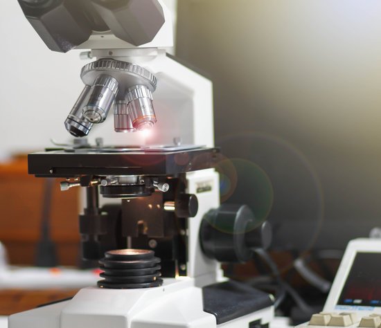
Which part of the microscope holds the objective lenses? Revolving Nosepiece or Turret: This is the part that holds two or more objective lenses and can be rotated to easily change power. Objective Lenses: Usually you will find 3 or 4 objective lenses on a microscope.
Which part of the microscope are objective? Nosepiece: The upper part of a compound microscope that holds the objective lens. Also called a revolving nosepiece or turret.
What are the objective lenses attached to? An optical microscope is used with multiple objectives attached to a part called revolving nosepiece. Commonly, multiple combined objectives with a different magnification are attached to this revolving nosepiece so as to smoothly change magnification from low to high only by revolving the nosepiece.
What information does SEM provide about the sample? A scanning electron microscope (SEM) is a type of electron microscope that produces images of a sample by scanning the surface with a focused beam of electrons. The electrons interact with atoms in the sample, producing various signals that contain information about the surface topography and composition of the sample.
Which part of the microscope holds the objective lenses? – Related Questions
What type of light is used for confocal microscope?
Similar to the widefield microscope, the confocal microscope uses fluorescence optics. Instead of illuminating the whole sample at once, laser light is focused onto a defined spot at a specific depth within the sample. This leads to the emission of fluorescent light at exactly this point.
How much is an electron microscope cost?
The price of a new electron microscope can range from $80,000 to $10,000,000 depending on certain configurations, customizations, components, and resolution, but the average cost of an electron microscope is $294,000. The price of electron microscopes can also vary by type of electron microscope.
Did robert hooke invented the compact microscope?
Although Hooke did not make his own microscopes, he was heavily involved with the overall design and optical characteristics. The microscopes were actually made by London instrument maker Christopher Cock, who enjoyed a great deal of success due to the popularity of this microscope design and Hooke’s book.
What is the condenser and control in microscope?
The Abbe condenser, which was originally designed for Zeiss, is mounted below the stage of the microscope. The condenser concentrates and controls the light that passes through the specimen prior to entering the objective.
Who was the individual that developed the first compound microscope?
A Dutch father-son team named Hans and Zacharias Janssen invented the first so-called compound microscope in the late 16th century when they discovered that, if they put a lens at the top and bottom of a tube and looked through it, objects on the other end became magnified.
What liquid do you use for microscope lenses?
Remove oily dirt using either a lens cleaning fluid or absolute ethanol on a cotton swab or lens tissue. Stubborn contamination may require several passes, or a stronger solvent such as methanol or acetone.
Why should a microscope slide and coverslip be held?
The main function of the cover slip is to keep solid specimens pressed flat, and liquid samples shaped into a flat layer of even thickness. This is necessary because high-resolution microscopes have a very narrow region within which they focus. The cover glass often has several other functions.
How is a microscope similar to a refracting telescope?
Telescopes and microscopes are both used to magnify images. However, a telescope looks at objects that are far away and help you see them as a larger image where a microscope looks at objects that are very small, but close up to enlarge them so you can see them.
Which microscope is best for viewing living organisms?
Light microscopes are advantageous for viewing living organisms, but since individual cells are generally transparent, their components are not distinguishable unless they are colored with special stains.
How does the letter e appear in a microscope?
The letter “e” appears upside down and backwards under a microscope. Either, diatoms are single celled, or they do not have a cell wall.
When was transmission electron microscopes invented?
Ernst Ruska at the University of Berlin, along with Max Knoll, combined these characteristics and built the first transmission electron microscope (TEM) in 1931, for which Ruska was awarded the Nobel Prize for Physics in 1986.
What moves the stage left and right on a microscope?
The stage clamp holds the microscope slide in place. Below the stage is a set of knobs called the STAGE ADJUSTMENT KNOBS. The top (larger) stage adjustment knob moves the stage vertically (towards you and away from you). The bottom (smaller) stage adjustment knob moves the stage horizontally (left/ right).
What are ocular lenses on a microscope?
The ocular lenses are the lenses closest to the eye and usually have a 10x magnification. Since light microscopes use binocular lenses there is a lens for each eye.
How to identify wbc under microscope?
Microscopy. Given that all white blood cells are over 5 micrometers in diameter, they are large enough to be seen using a typical optical microscope (compound microscope). Staining with Leishman’s stain makes it possible to not only easily identify different types of leukocytes, but also count them.
What are some limitations of microscopic observation?
The microscope can’t produce the image of an object that is smaller than the length of the light wave. Any object that’s less than half the wavelength of the microscope’s illumination source is not visible under that microscope. Light microscopes use visible light.
What cellular structures can be seen with a light microscope?
Note: The nucleus, cytoplasm, cell membrane, chloroplasts and cell wall are organelles which can be seen under a light microscope.
Which is the high power lens on a microscope?
The high-powered objective lens (also called “high dry” lens) is ideal for observing fine details within a specimen sample. The total magnification of a high-power objective lens combined with a 10x eyepiece is equal to 400x magnification, giving you a very detailed picture of the specimen in your slide.
What power microscope to see tardigrades?
In the right light you can actually see them with the naked eye. But researchers who work with tardigrades see them as they appear through a dissecting microscope of 20- to 30-power magnification—as charismatic miniature animals. Most tiny invertebrates dart about frantically.
What organism is unicellular and microscopic?
Most unicellular organisms are of microscopic size and are thus classified as microorganisms. However, some unicellular protists and bacteria are macroscopic and visible to the naked eye.

