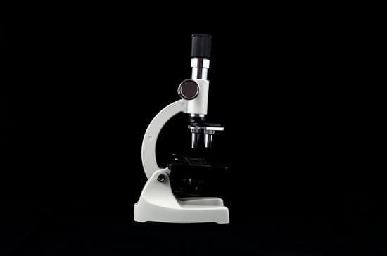
Which types of microscopes can produce 3d images? In scanning electron microscopy (SEM), a beam of electrons moves back and forth across the surface of a cell or tissue, creating a detailed image of the 3D surface.
What type of microscope is used in forensic science? Electron microscopy (EM) has a wide variety of applications in forensic investigation. Numerous crime-scene micro-traces, including glass and paint fragments, tool marks, drugs, explosives and gunshot residue (GSR) can be visually and chemically analyzed with scanning electron microscopy (SEM).
How do microscopes analyze evidence? These tools are used to characterize forensic evidence like fabrics, metals, textile or glass. Microscopic imaging can also aid in identifying scratches and indents from tool marks, blood, hair classification, particle analysis, or scrutinizing residues such as sand, mud, and diatoms.
What is the most common microscope used in a forensic laboratory? The stereoscopic microscope is the most frequently used and versatile microscope found in the crime laboratory. When you increase the compound microscope magnification its field of view decreases.
Which types of microscopes can produce 3d images? – Related Questions
What is the highest magnification of a compound light microscope?
On a stock, high-performance compound light microscope, magnification levels of 1000x can be achieved (10x ocular lens, 100x objective lens). With that said, the maximum magnification level of a light microscope at the high end of the performance spectrum is 2000x magnification (20x ocular, 100x objective).
Who invented microscope first time when and where?
It’s not clear who invented the first microscope, but the Dutch spectacle maker Zacharias Janssen (b. 1585) is credited with making one of the earliest compound microscopes (ones that used two lenses) around 1600. The earliest microscopes could magnify an object up to 20 or 30 times its normal size.
What cell structures can you see with a light microscope?
Note: The nucleus, cytoplasm, cell membrane, chloroplasts and cell wall are organelles which can be seen under a light microscope.
How to adjust ocular lens on microscope?
This is very simple – most microscopes have an adjuster wheel in the centre of the eyepieces to adjust the distance. Otherwise, slide the eyepiece housing to match the width of your eyes. Once you have set this distance, you can then make the diopter adjustment.
What can be seen only with an electron microscope?
Mitochondria are visible with the light microscope but can’t be seen in detail. Ribosomes are only visible with the electron microscope.
Can any microscope see atoms?
Atoms are really small. So small, in fact, that it’s impossible to see one with the naked eye, even with the most powerful of microscopes. … Now, a photograph shows a single atom floating in an electric field, and it’s large enough to see without any kind of microscope.
Which microscope has the magnification of 400x?
The compound microscope typically has three or four magnifications – 40x, 100x, 400x, and sometimes 1000x. At 40x magnification you will be able to see 5mm. At 100x magnification you will be able to see 2mm. At 400x magnification you will be able to see 0.45mm, or 450 microns.
How does the first microscope work?
A Dutch father-son team named Hans and Zacharias Janssen invented the first so-called compound microscope in the late 16th century when they discovered that, if they put a lens at the top and bottom of a tube and looked through it, objects on the other end became magnified.
How is something viewed with a microscope?
It is through the microscope’s lenses that the image of an object can be magnified and observed in detail. … When light reflects off of an object being viewed under the microscope and passes through the lens, it bends towards the eye. This makes the object look bigger than it actually is.
Why are binocular microscopes necessary for microscopy?
Binocular microscopes have two eye pieces, which can make it easier for the viewer to observe slide samples. Many users also find binocular microscopes to be more comfortable to use instead of the monocular microscopes.
What microscope magnification to see bacteria in water?
In order to actually see bacteria swimming, you’ll need a lens with at least a 400x magnification. A 1000x magnification can show bacteria in stunning detail. However, at a higher magnification, it can be increasingly difficult to keep them in focus as they move.
What does a filter on a microscope do?
Microscopy filters are used to filter out specific wavelengths of light thereby increasing contrast, blocking ambient light, removing IR or UV radiation. Filters are generally fitted over the illuminating device below the iris diaphragm.
What is the importance of electron microscope?
Electron microscopes are important for the depth of detail they show, which has led to a variety of important discoveries. Understanding their importance requires an understanding of how they work, and how this has led to further discovery.
How to calculate field diameter of a microscope?
The field size or diameter at a given magnification is calculated as the field number divided by the objective magnification. If the ×40 objective is used, the diameter of the field of view becomes 20 mm/40 (compared with no objective) or 0.5 mm.
How to use a compound microscope for beginners?
Turn the revolving turret (2) so that the lowest power objective lens (eg. 4x) is clicked into position. Place the microscope slide on the stage (6) and fasten it with the stage clips. Look at the objective lens (3) and the stage from the side and turn the focus knob (4) so the stage moves upward.
When would you use a sem microscope?
In general, if you need to look at a relatively large area and only need surface details, SEM is ideal. If you need internal details of small samples at near-atomic resolution, TEM will be necessary.
What role does the microscope play in cell theory?
Explanation: With the development and improvement of the light microscope, the theory created by Sir Robert Hooke that organisms would be made of cells was confirmed as scientist were able to actually see cells in tissues placed under the microscope.
What is meant by the magnification of a microscope?
Magnification is the ability of a microscope to produce an image of an object at a scale larger (or even smaller) than its actual size.
How to identify malaria parasites under the microscope?
Malaria parasites can be identified by examining under the microscope a drop of the patient’s blood, spread out as a “blood smear” on a microscope slide. Prior to examination, the specimen is stained (most often with the Giemsa stain) to give the parasites a distinctive appearance.
What you should use when carrying a microscope?
Always keep your microscope covered when not in use. Always carry a microscope with both hands. Grasp the arm with one hand and place the other hand under the base for support.

