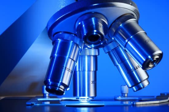
Who discovered cells under a microscope? Initially discovered by Robert Hooke in 1665, the cell has a rich and interesting history that has ultimately given way to many of today’s scientific advancements.
What microscope originally discovered cells? Called the electron microscope, it used a beam of electrons instead of light to observe extremely small objects. With an electron microscope, scientists could finally see the tiny structures inside cells. In fact, they could even see individual molecules and atoms. The electron microscope had a huge impact on biology.
What did Robert Hooke discover about cells? While observing cork through his microscope, Hooke saw tiny boxlike cavities, which he illustrated and described as cells. He had discovered plant cells! Hooke’s discovery led to the understanding of cells as the smallest units of life—the foundation of cell theory.
Who are the 10 scientists who discovered cells? Landmarks in Discovery of Cells
Who discovered cells under a microscope? – Related Questions
How did the invention of microscopes help scientists understand cells?
Microscopes allow humans to see cells that are too tiny to see with the naked eye. Therefore, once they were invented, a whole new microscopic world emerged for people to discover. … It allowed them to observe Eukaryotic cells with a nucleus and membrane-bound organelles that perform different life functions.
When was the first electron microscope developed?
Ernst Ruska at the University of Berlin, along with Max Knoll, combined these characteristics and built the first transmission electron microscope (TEM) in 1931, for which Ruska was awarded the Nobel Prize for Physics in 1986.
Why the atomic force microscope is important to scientists?
AFM is a very powerful technique for the biological sciences, allowing samples to be imaged in situ in physiological conditions. … High-resolution imaging, therefore, allows molecular scale features to be identified in the native environment of the sample and in real time.
Can you see a human skin cell without a microscope?
You can see the tissue they form (example: skin) but you cannot visualize them without use of microscope.
How to add magnification of a microscope?
Total Magnification: To figure the total magnification of an image that you are viewing through the microscope is really quite simple. To get the total magnification take the power of the objective (4X, 10X, 40x) and multiply by the power of the eyepiece, usually 10X.
Will phones replace microscopes?
Smartphones could never replace sophisticated laboratory microscopes. Yet, they can be good enough to make learning and diagnosing more accessible. Especially where technology and economy prevent professionals from acquiring and using advanced devices. Mobile phones have another advantage.
What is a transmission light microscope?
Definition. Transmission light microscopy is any optical microscopy technique that relies on detection of light from the opposite side of the sample that was illuminated.
Who invented of the microscope in 1666?
Antoni Van Leeuwenhoek (1635-1723) was a Dutch tradesman who became interested in microscopy while on a visit to London in 1666. Returning home, he began making simple microscopes of the sort that Robert Hooke had described in his, Micrographia, and using them to discover objects invisible to the naked eye.
What did leeuwenhoek see with his microscope?
The van Leeuwenhoek microscope provided man with the first glimpse of bacteria. In 1674, van Leeuwenhoek first described seeing red blood cells. Crystals, spermatozoa, fish ova, salt, leaf veins, and muscle cell were seen and detailed by him.
Where is the ocular lens located on a microscope?
Enlargement or magnification of a specimen is the function of a two-lens system; the ocular lens is found in the eyepiece, and the objective lens is situated in a revolving nose-piece. These lenses are separated by the body tube.
Why are light microscopes limited to 1000x magnification?
The maximum magnification power of optical microscopes is typically limited to around 1000x because of the limited resolving power of visible light. … Modified environments such as the use of oil or ultraviolet light can increase the magnification.
What is the microscopic structure of skeletal muscle?
Skeletal muscle fibers are long, multinucleated cells. The membrane of the cell is the sarcolemma; the cytoplasm of the cell is the sarcoplasm. The sarcoplasmic reticulum (SR) is a form of endoplasmic reticulum. Muscle fibers are composed of myofibrils which are composed of sarcomeres linked in series.
Why we use a microscope?
A microscope is an instrument that is used to magnify small objects. Some microscopes can even be used to observe an object at the cellular level, allowing scientists to see the shape of a cell, its nucleus, mitochondria, and other organelles.
Can we see viruses with a light microscope?
Standard light microscopes allow us to see our cells clearly. However, these microscopes are limited by light itself as they cannot show anything smaller than half the wavelength of visible light – and viruses are much smaller than this.
How good are digital microscopes?
The digital version is great for speed, convenience, and high-quality images that need to be taken multiple times. The optical microscope is great if you don’t need any of the fancy hardware to get your job done. … That said, both microscopes can be great to have on hand and it can be beneficial to use both.
What is magnification of simple microscope?
1 + D/F, where D is the least distance of distinct vision, and F is the focal length of the convex lens.
What is the resolution of scanning electron microscope?
Scanning electron microscope (SEM) is one of the most widely used instrumental methods for the examination and analysis of micro- and nanoparticle imaging characterization of solid objects. One of the reasons that SEM is preferred for particle size analysis is due to its resolution of 10 nm, that is, 100 Å.
What is a aperture on a microscope?
Numerical Aperture and Resolution. The numerical aperture of a microscope objective is the measure of its ability to gather light and to resolve fine specimen detail while working at a fixed object (or specimen) distance. … The smaller the object, the more pronounced the diffraction of incident light rays will be.
How to use a micrometer microscope?
Procedure. Place a stage micrometer on the microscope stage, and using the lowest magnification (4X), focus on the grid of the stage micrometer. Rotate the ocular micrometer by turning the appropriate eyepiece. Move the stage until you superimpose the lines of the ocular micrometer upon those of the stage micrometer.
How to microscopes help us?
A microscope lets the user see the tiniest parts of our world: microbes, small structures within larger objects and even the molecules that are the building blocks of all matter. The ability to see otherwise invisible things enriches our lives on many levels.
What is the vegetable that has microscopic worms in it?
While most veggies have them, cabbage and cauliflower are specially notorious of harboring them. These worms are so small that they cannot be seen with naked eyes as they are hidden inside the layers of cabbage or cauliflower.

