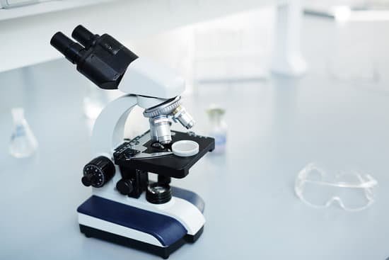
Who discovered microscope wiki? Several revolve around the spectacle-making centers in the Netherlands, including claims it was invented in 1590 by Zacharias Janssen (claim made by his son) or Zacharias’ father, Hans Martens, or both, claims it was invented by their neighbor and rival spectacle maker, Hans Lippershey (who applied for the first …
What are the 5 uses of microscope? They are used in different fields for different purposes. Some of their uses are tissue analysis, the examination of forensic evidence, to determine the health of the ecosystem, studying the role of protein within the cell, and the study of atomic structure.
What are four uses of a microscope? Importance of Microscope in our Daily Life. Microscopes have opened up many doors in science. … Microscopes are not just used to observe cells and their structure but are also used in many industries. For example, electron microscopes help create and observe extremely tiny electrical circuits found on Silicon microchips.
What is the resolution of a scanning electron microscope? Scanning electron microscope (SEM) is one of the most widely used instrumental methods for the examination and analysis of micro- and nanoparticle imaging characterization of solid objects. One of the reasons that SEM is preferred for particle size analysis is due to its resolution of 10 nm, that is, 100 Å.
Who discovered microscope wiki? – Related Questions
Were cells discovered with the electron microscope?
Then, in the 1950s, a new type of the microscope was invented. Called the electron microscope, it used a beam of electrons instead of light to observe extremely small objects. With an electron microscope, scientists could finally see the tiny structures inside cells.
How microscopic is sperm?
The air-fixed, stained spermatozoa are observed under a bright-light microscope at 400x or 1000x magnification. Their viability and mor- phology can be analysed at the same time. Those appearing red-pink in colour have a damaged membrane whereas white sperm are viable, as in Photo 2.
When should i use a transmission electron microscope?
Transmission electron microscopy is a major analytical method in the physical, chemical and biological sciences. TEMs find application in cancer research, virology, and materials science as well as pollution, nanotechnology and semiconductor research, but also in other fields such as paleontology and palynology.
Can you see a female egg without a microscope?
Most cells aren’t visible to the naked eye: you need a microscope to see them. The human egg cell is an exception, it’s actually the biggest cell in the body and can be seen without a microscope.
What does sickle cell anemia look like under a microscope?
Healthy red blood cells are round and move easily through even the smallest blood vessels to deliver oxygen to all parts of the body. In someone with SCD, red blood cells become hard and sticky. Viewed under a microscope they appear C-shaped, like the farm tool called a “sickle.”
What things can be seen with a light microscope?
Explanation: You can see most bacteria and some organelles like mitochondria plus the human egg. You can not see the very smallest bacteria, viruses, macromolecules, ribosomes, proteins, and of course atoms.
Who developed microscopes?
The development of the microscope allowed scientists to make new insights into the body and disease. It’s not clear who invented the first microscope, but the Dutch spectacle maker Zacharias Janssen (b. 1585) is credited with making one of the earliest compound microscopes (ones that used two lenses) around 1600.
What is microscopic examination of living tissue?
Biopsy – The removal and examination, usually microscopic, of tissue from the living body, performed to establish precise diagnosis.
What are the two different types of slides on microscope?
You will be using two main types of slides, 1) the common flat glass slide, and 2) the depression or well slides. Well slides have a small well, or indentation, in the center to hold a drop of water or liquid substance. They are more expensive and usually used without a cover slip.
Why is the microscopic examination of urine a ppm procedure?
PPM procedures are a select group of moderately complex microscopic tests that do not meet the criteria for waiver because they are not simple procedures; they require training and specific skills for test performance and they must meet certain other criteria.
What is the cost of a transmission electron microscope?
The cost of a transmission electron microscope (TEM) can range from $300,000 to $10,000,000. The cost of a focused ion beam electron microscope (FIB) can range from $500,000 to $4,000,000.
What is the total magnification of a microscope lens?
Total Magnification: To figure the total magnification of an image that you are viewing through the microscope is really quite simple. To get the total magnification take the power of the objective (4X, 10X, 40x) and multiply by the power of the eyepiece, usually 10X.
What is the difference between microscope and magnifying glass?
One difference between a magnifying glass and a compound light microscope is that a magnifying glass uses one lens to magnify an object while a compound microscope uses two or more lenses.
What microscope is used to see viruses in infected cells?
Electron microscopy (EM) has long been used in the discovery and description of viruses. Organisms smaller than bacteria have been known to exist since the late 19th century (11), but the first EM visualization of a virus came only after the electron microscope was developed.
What do we use microscopes for?
A microscope is an instrument that is used to magnify small objects. Some microscopes can even be used to observe an object at the cellular level, allowing scientists to see the shape of a cell, its nucleus, mitochondria, and other organelles.
How to use compound microscopes?
Turn the revolving turret (2) so that the lowest power objective lens (eg. 4x) is clicked into position. Place the microscope slide on the stage (6) and fasten it with the stage clips. Look at the objective lens (3) and the stage from the side and turn the focus knob (4) so the stage moves upward.
How to increase microscope resolution?
To achieve the maximum (theoretical) resolution in a microscope system, each of the optical components should be of the highest NA available (taking into consideration the angular aperture). In addition, using a shorter wavelength of light to view the specimen will increase the resolution.
What is a c mount microscope?
A C mount is a type of lens mount commonly found on 16 mm movie cameras, closed-circuit television cameras, machine vision cameras and microscope phototubes. C-mount lenses provide a male thread, which mates with a female thread on the camera. … The flange focal distance is 17.526 millimetres (0.6900 in) for a C mount.

