Who invented microscope wikipedia? Antonie van Leeuwenhoek (1632–1724) is credited with bringing the microscope to the attention of biologists, even though simple magnifying lenses were already being produced in the 16th century. Van Leeuwenhoek’s home-made microscopes were simple microscopes, with a single very small, yet strong lens.
Who first invented the microscope? The development of the microscope allowed scientists to make new insights into the body and disease. It’s not clear who invented the first microscope, but the Dutch spectacle maker Zacharias Janssen (b. 1585) is credited with making one of the earliest compound microscopes (ones that used two lenses) around 1600.
Who invented the first microscope Wikipedia?
Who invented microscope and in what year? 1590: Two Dutch spectacle-makers and father-and-son team, Hans and Zacharias Janssen, create the first microscope.
Who invented microscope wikipedia? – Related Questions
Why are electron microscope images in black and white?
Why do electron microscopes produce black and white images? The reason is pretty basic: color is a property of light (i.e., photons), and since electron microscopes use an electron beam to image a specimen, there’s no color information recorded. … However, the images it produces contain only two colors – red and green.
How microscope magnification works?
In simple magnification, light from an object passes through a biconvex lens and is bent (refracted) towards your eye. … Both of these contribute to the magnification of the object. The eyepiece lens usually magnifies 10x, and a typical objective lens magnifies 40x.
What does asymptomatic microscopic hematuria mean?
Asymptomatic microscopic hematuria is an important clinical sign for urinary tract malignancy. Risk factors for urinary tract malignancy include being male, being older, being a past or current smoker, having gross hematuria, and having a history of pelvic irradiation.
What type of microscope is a tem?
Transmission electron microscopes (TEM) are microscopes that use a particle beam of electrons to visualize specimens and generate a highly-magnified image. TEMs can magnify objects up to 2 million times.
How much for a cheap microscope?
With such brands as Amscope and Omax, users will be surprised by the good level of quality they can get for microscopes that cost less than $300. The following are a few examples of low cost good quality microscopes from Amscope and Omax.
Can’t you see an element through a microscope?
Atoms are really small. So small, in fact, that it’s impossible to see one with the naked eye, even with the most powerful of microscopes. At least, that used to be true. Now, a photograph shows a single atom floating in an electric field, and it’s large enough to see without any kind of microscope.
How much lenses does a compound microscope have?
A compound microscope is an upright microscope that uses two sets of lenses (a compound lens system) to obtain higher magnification than a stereo microscope. A compound microscope provides a two-dimensional image, while a stereo microscope provides a three-dimensional image.
What is a hot stage microscope used for?
Hot stage microscopy is a powerful method which is widely used to visually examine all kinds of thermal transitions. In the HS82, samples are heated or cooled while they are observed under the microscope. The HS84 DSC hot-stage can even simultaneously measure heat flow.
How does a monocular light microscope work?
The resolution limit is set by the wave nature of light at approximately 1000 times magnification. The microscope in the cabinet uses an Abbe condenser lens. … A condenser lens sits underneath the specimen and above the illumination source and concentrates the light through the specimen and into the objective lens.
What is the function of condenser in compound microscope?
On upright microscopes, the condenser is located beneath the stage and serves to gather wavefronts from the microscope light source and concentrate them into a cone of light that illuminates the specimen with uniform intensity over the entire viewfield.
Where was the first compound microscope invented?
Janssen was the son of a spectacle maker named Hans Janssen, in Middleburg, Holland, and while Zacharias is credited with inventing the compound microscope, most historians surmise that his father must have played a vital role, since Zacharias was still in his teens in the 1590s.
How to focus compound light microscope?
Look at the objective lens (3) and the stage from the side and turn the focus knob (4) so the stage moves upward. Move it up as far as it will go without letting the objective touch the coverslip. Look through the eyepiece (1) and move the focus knob until the image comes into focus.
What does the nosepiece do on a microscope?
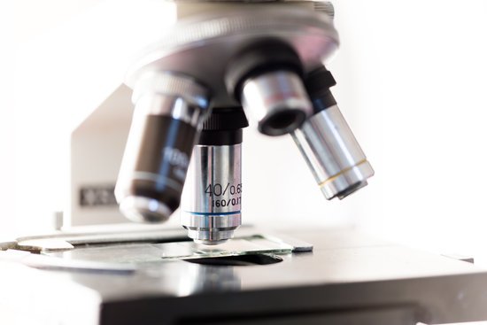
Nosepiece houses the objectives. The objectives are exposed and are mounted on a rotating turret so that different objectives can be conveniently selected. Standard objectives include 4x, 10x, 40x and 100x although different power objectives are available. Coarse and Fine Focus knobs are used to focus the microscope.
What is the source of light in electron microscope?
An electron microscope is a microscope that uses a beam of accelerated electrons as a source of illumination.
How many times can a transmission electron microscope magnify?
TEM: magnifies 50 to ~50 million times; the specimen appears flat. SEM: magnifies 5 to ~ 500,000 times; sharp images of surface features. STEM: magnifies 5 to ~50 million times; the specimen appears flat.
Who created the transmission electron microscope?
Ernst Ruska at the University of Berlin, along with Max Knoll, combined these characteristics and built the first transmission electron microscope (TEM) in 1931, for which Ruska was awarded the Nobel Prize for Physics in 1986.
How do you find total magnification of a compound microscope?
The total magnification is calculated by multiplying the magnification of the ocular lens by the magnification of the objective lens.
Can dehydration cause microscopic blood in urine?
For example, not getting enough fluids (dehydration), taking certain medicines, or having a liver problem can change the colour of your urine. Eating foods such as beets, rhubarb, or blackberries or foods with red food colouring can make your urine look red or pink.
What are standard microscope powers?
They almost always consist of 4x, 10x, 40x and 100x powers. When coupled with a 10x (most common) eyepiece lens, total magnification is 40x (4x times 10x), 100x , 400x and 1000x. To have good resolution at 1000x, you will need a relatively sophisticated microscope with an Abbe condenser.
Can we see dna without a microscope?
Many people assume that because DNA is so small, we can’t see it without powerful microscopes. But in fact, DNA can be easily seen with the naked eye when collected from thousands of cells.
Does microscopic colitis cause nausea?
Microscopic colitis is a condition of the colon that causes watery diarrhea, pain, and nausea. It is more common among people older than 60 years of age. Budesonide is the most effective treatment and has a low risk of side effects. However, the cost of the drug may be an issue for some patients.

