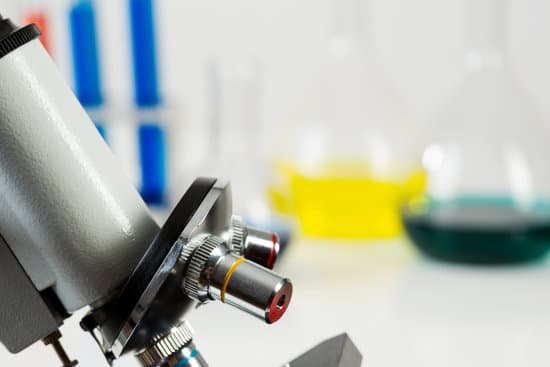
Why are microscopes important tools in the field of biology? The microscope is important because biology mainly deals with the study of cells (and their contents), genes, and all organisms. Some organisms are so small that they can only be seen by using magnifications of ×2000−×25000 , which can only be achieved by a microscope.
What is the importance of microscope as a tool in the field of science? The invention of the microscope allowed scientists to see cells, bacteria, and many other structures that are too small to be seen with the unaided eye. It gave them a direct view into the unseen world of the extremely tiny. You can get a glimpse of that world in Figure below.
How are microscopes related to biology? Light microscopy is one of the least invasive techniques used to access information from various biological scales in living cells. The combination of molecular biology and imaging provides a bottom-up tool for direct insight into how molecular processes work on a cellular scale.
How do you switch objectives on a microscope? When focusing on a slide, ALWAYS start with either the 4X or 10X objective. Once you have the object in focus, then switch to the next higher power objective. Re-focus on the image and then switch to the next highest power.
Why are microscopes important tools in the field of biology? – Related Questions
Who found light microscope?
The Dutch spectacle maker Hans Janssen and his son Zacharias are generally credited with creating these compound microscopes. The two of them built what was probably the first compound microscope in the last decade of the 16th century.
How to check microscope for germs?
Viewing bacteria under a microscope is much the same as looking at anything under a microscope. Prepare the sample of bacteria on a slide and place under the microscope on the stage. Adjust the focus then change the objective lens until the bacteria come into the field of view.
What does moderate microscopic blood in urine mean?
Microscopic urinary bleeding is a common symptom of glomerulonephritis, an inflammation of the kidneys’ filtering system. Glomerulonephritis may be part of a systemic disease, such as diabetes, or it can occur on its own.
Why is helium used for microscopes?
Helium ions have shorter wavelengths than electrons, so helium ions can form a more tightly focused beam. For a microscope, that means better image resolution. … The helium-derived images have higher surface contrast and better depth of field, so more of the image is in focus than in SEM-derived images.
Which part of microscope used to focus coarse adjustment?
Coarse Adjustment Knob- The coarse adjustment knob located on the arm of the microscope moves the stage up and down to bring the specimen into focus. The gearing mechanism of the adjustment produces a large vertical movement of the stage with only a partial revolution of the knob.
Do latex condoms have microscopic holes?
Condoms used to be made of natural skin (including lambskin) or of rubber. That’s why they are called “rubbers.” Most condoms today are made of latex. Lambskin condoms can prevent pregnancy. However, they have tiny holes (pores) that are large enough for HIV to get through.
How many types of microscope name them?
There are several different types of microscopes used in light microscopy, and the four most popular types are Compound, Stereo, Digital and the Pocket or handheld microscopes.
Are digital microscopes any good?
The digital version is great for speed, convenience, and high-quality images that need to be taken multiple times. The optical microscope is great if you don’t need any of the fancy hardware to get your job done.
Which branch of microscopic anatomy is the study of tissues?
Histology, also known as microscopic anatomy or microanatomy, is the branch of biology which studies the microscopic anatomy of biological tissues. Histology is the microscopic counterpart to gross anatomy, which looks at larger structures visible without a microscope.
How to find out the total magnification of a microscope?
To figure the total magnification of an image that you are viewing through the microscope is really quite simple. To get the total magnification take the power of the objective (4X, 10X, 40x) and multiply by the power of the eyepiece, usually 10X.
What branch of microscopic anatomy is the study of tissues?
Histology, also known as microscopic anatomy or microanatomy, is the branch of biology which studies the microscopic anatomy of biological tissues. Histology is the microscopic counterpart to gross anatomy, which looks at larger structures visible without a microscope.
What are portable microscopes used for?
Applications include use by emergency medical technicians, trauma and emergency room practitioners, field study by scientists and hobbyists. Manufacturing companies find portable microscopes invaluable for finding imperfections in electrical components, metals, optics, glassware and structural defects on machinery.
Why need a color wheel on microscope?
How is it used? Filters are used to increase contrast and color correction for visual observations of specimens or slides. Simply turn the wheel to put the filter you need into the path of light from your condenser-illuminator. …
What kind of microscope is needed to see blood?
The compound microscope can be used to view a variety of samples, some of which include: blood cells, cheek cells, parasites, bacteria, algae, tissue, and thin sections of organs. Compound microscopes are used to view samples that can not be seen with the naked eye.
How much can electron microscopes magnify images?
This makes electron microscopes more powerful than light microscopes. A light microscope can magnify things up to 2000x, but an electron microscope can magnify between 1 and 50 million times depending on which type you use! To see the results, look at the image below.
Which lens should face the stage when storing the microscope?
Always place the 4X objective over the stage and be sure the stage is at its lowest position before putting the microscope away. 9. Always turn off the light before putting the microscope away. 10.
What is microscope ergonomics?
Microscope ergonomics is a priority for clinical routine microscopy. … Ergonomic microscopes are characterized by flexible headpieces; lowered buttons and controls help the operator to maintain a comfortable posture. A combination of equipment, setup and customisation minimises the risk of injury.
What does a microscope do the image you are viewing?
A microscope is an instrument that can be used to observe small objects, even cells. The image of an object is magnified through at least one lens in the microscope. This lens bends light toward the eye and makes an object appear larger than it actually is.
What are the illuminating parts of microscope?
It consists of mainly three parts: Mechanical part – base, c-shaped arm and stage. Magnifying part – objective lens and ocular lens. Illuminating part – sub stage condenser, iris diaphragm, light source.

