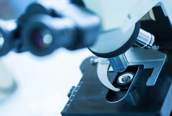
Why couldn’t scientists identify spanish flu with optical microscope? Unlike many bacteria, it was too small to be seen through an optical microscope. Without having actually seen viruses, scientists debated their nature: were they organism or toxin, liquid or particle, dead or alive? They were veiled in mystery, and nobody suspected that they could be the cause of flu.
What is the importance of microscope in scientific? The invention of the microscope allowed scientists to see cells, bacteria, and many other structures that are too small to be seen with the unaided eye. It gave them a direct view into the unseen world of the extremely tiny. You can get a glimpse of that world in Figure below.
Why is the microscope important? The microscope is important because biology mainly deals with the study of cells (and their contents), genes, and all organisms. Some organisms are so small that they can only be seen by using magnifications of ×2000−×25000 , which can only be achieved by a microscope. Cells are too small to be seen with the naked eye.
What is the importance of using microscope for you as a student? Many of us probably used a microscope or stereoscope in high school or even before. These instruments enable students to observe very small structural details that are hard to see by eye, such as the structure of smooth muscle, cellular division, or the details of an insect.
Why couldn’t scientists identify spanish flu with optical microscope? – Related Questions
How to remove air bubbles from microscope slide?
Apply a vacuum: You can remove air bubbles by placing the slide into a vacuum. THe bubbles will expand and move out beneath the cover glass. Dehydrate the specimen: Place the specimen into alcohol. Some specimens will shrink and lose water and air.
What microscope do you use for colocalization?
Confocal microscopy is used to test whether two fluorescently labeled molecules are associated with one another. If one is labeled red and the other is labeled green, an image that combines them may show a yellow color where the red and green pixels overlap.
Which microscope produces three dimensional images?
The two microscopes that are able to produce a three- dimensional image of the object are scanning tunnel microscope and transmission electron microscope.
What era was the microscope invented?
In the late 16th century several Dutch lens makers designed devices that magnified objects, but in 1609 Galileo Galilei perfected the first device known as a microscope.
Why use coverslip on microscope?
When viewing any slide with a microscope, a small square or circle of thin glass called a coverslip is placed over the specimen. It protects the microscope and prevents the slide from drying out when it’s being examined. The coverslip is lowered gently onto the specimen using a mounted needle .
What is f 200 microscope?
JEM-F200 is a new field emission transmission electron microscope, which features higher spatial resolution and analytical performance, an easy to use new operation system for multi-purpose operation, a smart appearance, and various environmentally friendly, energy saving system.
Can you analyze blood through a microscope?
A blood smear is a sample of blood that’s tested on a specially treated slide. For a blood smear test, a laboratory professional examines the slide under a microscope and looks at the size, shape, and number of different types of blood cells.
How many types of compound microscope?
A compound microscope can come in several types such as biological microscopes, polarizing microscopes, phase contrast microscopes, or florescence microscopes with uses varying for each.
What are the 4 types of microscopes and their functions?
There are several different types of microscopes used in light microscopy, and the four most popular types are Compound, Stereo, Digital and the Pocket or handheld microscopes. Some types are best suited for biological applications, where others are best for classroom or personal hobby use.
Why the light microscope is also called the compound microscope?
The compound light microscope is a tool containing two lenses, which magnify, and a variety of knobs used to move and focus the specimen. Since it uses more than one lens, it is sometimes called the compound microscope in addition to being referred to as being a light microscope.
What is microscopic metastatic disease?
Micrometastases are a small collection of cancer cells that have been shed from the original tumor and spread to another part of the body through the blood or lymph nodes.
What is the function of oil immersion on a microscope?
Immersion oil increases the resolving power of the microscope by replacing the air gap between the immersion objective lens and cover glass with a high refractive index medium and reducing light refraction.
How to make a microscope parfocal?
1. Focus on the specimen using the focusing knobs until you get a sharp image through the monitor / display. 2.
What is the minimum objective magnification of a microscope?
A scanning objective lens provides the lowest magnification power of all objective lenses. 4x is a common magnification for scanning objectives and, when combined with the magnification power of a 10x eyepiece lens, a 4x scanning objective lens gives a total magnification of 40x.
Can a transmission electron microscope see wet samples?
Electron Microscopy (EM) is a prime tool for high-resolution imaging, which has been the cornerstone of our understanding of living organisms and our material environment. Because EM requires samples to be placed in a vacuum, it does not lend itself for use with wet samples.
Can chlamydia cause microscopic hematuria?
Specifically, the STDs that most commonly cause blood in urine are chlamydia and gonorrhea. Seeing blood in your urine can be very worrisome and the best course of action is to see a doctor if this symptom persists for several days.
What is a ua dipstick w reflex microscopic?
Urinalysis with Reflex to Microscopic – Dipstick urinalysis measures chemical constituents of urine. Microscopic examination helps to detect the presence of cells, bacteria, yeast and other formed elements.
Does the scanning electron microscope kill the specimen?
Preparation of a specimen for viewing under an electron microscope will kill it; therefore, live cells cannot be viewed using this type of microscopy. In addition, the electron beam moves best in a vacuum, making it impossible to view living materials.
What does microscopes kid mean?
Say: my-kro-skope. A microscope is a very powerful magnifying glass. The entire world – our bodies included – are made up of billions of tiny living things that are so small you can’t see them with just your eyes. But with a microscope, it’s possible to examine the cells of your body or a drop of blood.
What is the use of compound microscope?
Typically, a compound microscope is used for viewing samples at high magnification (40 – 1000x), which is achieved by the combined effect of two sets of lenses: the ocular lens (in the eyepiece) and the objective lenses (close to the sample).

