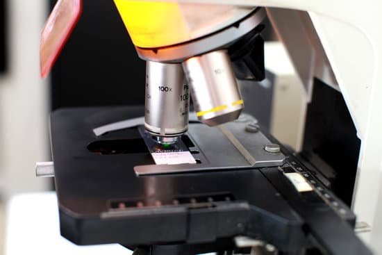
Why do electron microscopes have greater magnification? The electrons are fired at the sample very fast. When electrons travel at speed they behave a bit like light, so we can use them to make an image. But because electrons have a smaller wavelength than visible light they can reveal very tiny details. This makes electron microscopes more powerful than light microscopes.
Do electron microscopes have higher magnification? It has much higher magnification or resolving power than a normal light microscope. Although modern electron microscopes can magnify objects up to two million times, they are still based upon Ruska’s prototype and his correlation between wavelength and resolution.
Why electron microscopes allow for greater magnification than compound microscopes? Because a compound microscope uses light, its resolution is limited to . … Electrons, however, have a much smaller wavelength, and therefore the total magnification of a scanning electron microscope is 200,000 times with a resolution of . 02 nanometer.
Which microscope has a higher magnification? Out of all types of microscopes, the electron microscope has the greatest capability in achieving high magnification and resolution levels, enabling us to look at things right down to each individual atom.
Why do electron microscopes have greater magnification? – Related Questions
What is the purpose of coarse adjustment on a microscope?
COARSE ADJUSTMENT KNOB — A rapid control which allows for quick focusing by moving the objective lens or stage up and down. It is used for initial focusing.
Can chloroplast be seen under a light microscope?
Chloroplasts are larger than mitochondria and can be seen more easily by light microscopy. Since they contain chlorophyll, which is green, chloroplasts can be seen without staining and are clearly visible within living plant cells. … These living plant cells are viewed by light microscopy.
How should one carry the microscope?
Always keep your microscope covered when not in use. Always carry a microscope with both hands. Grasp the arm with one hand and place the other hand under the base for support.
What is this microscope used for?
A microscope is an instrument that can be used to observe small objects, even cells. The image of an object is magnified through at least one lens in the microscope. This lens bends light toward the eye and makes an object appear larger than it actually is.
Which microscope was invented by calvin goddard?
The comparison microscope was invented in the 1920s by American Army Colonel Calvin Goddard (1891–1955) who was working for the Bureau of Forensic Ballistics of the City of New York.
Who first discovered cells using a microscope?
The cell was first discovered by Robert Hooke in 1665 using a microscope. The first cell theory is credited to the work of Theodor Schwann and Matthias Jakob Schleiden in the 1830s.
What is the function of microscope slide?
A microscope slide is a thin flat piece of glass, typically 75 by 26 mm (3 by 1 inches) and about 1 mm thick, used to hold objects for examination under a microscope. Typically the object is mounted (secured) on the slide, and then both are inserted together in the microscope for viewing.
What is the scanning and tunneling electron microscope used for?
A scanning tunneling microscope (STM) is a type of microscope used for imaging surfaces at the atomic level. Its development in 1981 earned its inventors, Gerd Binnig and Heinrich Rohrer, then at IBM Zürich, the Nobel Prize in Physics in 1986. … This means that individual atoms can routinely be imaged and manipulated.
What is the use of microscope in lab?
The goal of any laboratory microscope is to produce clear, high-quality images, whether an optical microscope, which uses light to generate the image, a scanning or transmission electron microscope (using electrons), or a scanning probe microscope (using a probe).
Which lens has the greatest magnification in a microscope?
The oil immersion objective lens provides the most powerful magnification, with a whopping magnification total of 1000x when combined with a 10x eyepiece.
When was the very first microscope invented?
Lens Crafters Circa 1590: Invention of the Microscope. Every major field of science has benefited from the use of some form of microscope, an invention that dates back to the late 16th century and a modest Dutch eyeglass maker named Zacharias Janssen.
How to see water in a microscope?
First, suck up a small amount of the water in the container with an eye dropper. Then, carefully release the water onto a microscope slide. Once the water is on the slide, use a slide cover slip to cover it. This will spread the water out into a thin layer over the slide.
What can electron microscopes see that light microscopes cannot?
Electron microscopes use a beam of electrons instead of beams or rays of light. Living cells cannot be observed using an electron microscope because samples are placed in a vacuum.
What microscopically is on the surface of a penny?
A research engineer with the Canadian Centre for Electron Microscopy at McMaster University has produced a microscopic three-dimensional flag, hidden within the surface of a Canadian penny. Invisible to the naked eye, the microscopic Canadian flag flies on a flagpole 1/100th the diameter of a human hair.
What is stage clip in microscope?
Stage Clips are used when there is no mechanical stage. The viewer is required to move the slide manually to view different sections of the specimen. Aperture is the hole in the stage through which the base (transmitted) light reaches the stage.
What type of light does a compound microscope use?
A compound light microscope has its own light source in its base. The incandescent light from the light source is reflected by a condenser lens beneath the specimen, and the light passes through the specimen, up to the objective lens, then the projector lens sends the magnified image onto the eyepiece.
What kind of microscope is required for yeast?
In general: Yeast counting: All you need for this is a microscope with a basic transmitted light source and enough magnification to resolve individual yeast cells. Almost any microscope with 100x to 200x magnification (more on how to determine this, below) and a light source will suffice.
What is found on the revolving nosepiece of a microscope?
Revolving Nosepiece or Turret: This is the part that holds two or more objective lenses and can be rotated to easily change power. Objective Lenses: Usually you will find 3 or 4 objective lenses on a microscope. They almost always consist of 4X, 10X, 40X and 100X powers.
What the rules of use to focus a microscope?
To focus a microscope, rotate to the lowest-power objective, and place your sample under the stage clips. Play with the magnification using the coarse adjustment knob and move your slide around until it is centered.
What german botanist viewed many plants under a microscope?
Matthias Jacob Schleiden studied microscopic plant structures. In his studies, he observed that the different parts of the plant organism are composed of cells or derivatives of cells.
What part of the microscope supports the slide?
Stage: The flat platform where you place your slides. Stage clips hold the slides in place. Revolving Nosepiece or Turret: This is the part that holds two or more objective lenses and can be rotated to easily change power.

