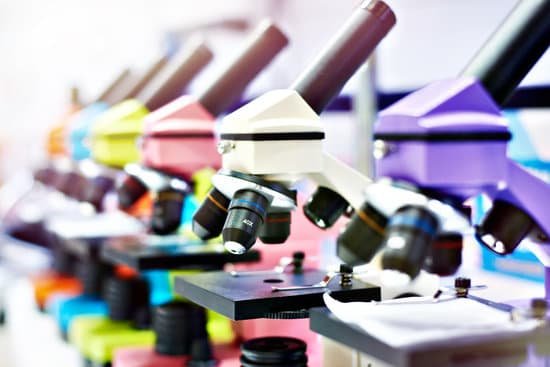
Why does an image flip in a microscope? An ocular lens is the one closest to your eye when looking through a microscope or telescope. … The objective lens is the lens that is closer to the object. The image will pass through the first lens and then the second lens, and because of the curvature of the first lens, the image will be inverted.
Does a microscope make an image upside down? A microscope is an instrument that magnifies an object. … The optics of a microscope’s lenses change the orientation of the image that the user sees. A specimen that is right-side up and facing right on the microscope slide will appear upside-down and facing left when viewed through a microscope, and vice versa.
What does it mean if the image is inverted when you look through the ocular lens? What does it mean that the image is inverted when you look through the ocular lenses? The ocular lens or eyepiece lens acts as a magnifying glass for the image, the ocular lens makes the light rays spread more so that they appear to come from a larger inverted image beyond the objective lands.
What are basophils stained with? Eosinophils (basic components that like acids) are dyed red by the acid stain, eosin. “Basophils” (acid that like base components) are dyed blue by the basic stain, hematoxylin.
Why does an image flip in a microscope? – Related Questions
What does a plant cell look like under a microscope?
Under the microscope, plant cells are seen as large rectangular interlocking blocks. The cell wall is distinctly visible around each cell. The cell wall is somewhat thick and is seen rightly when stained. The cytoplasm is also lightly stained containing a darkly stained nucleus at the periphery of the cell.
What year did zacharias janssen invent the microscope?
Lens Crafters Circa 1590: Invention of the Microscope. Every major field of science has benefited from the use of some form of microscope, an invention that dates back to the late 16th century and a modest Dutch eyeglass maker named Zacharias Janssen.
What is the light source on the microscope called?
Illuminator is the light source for a microscope, typically located in the base of the microscope. Most light microscopes use low voltage, halogen bulbs with continuous variable lighting control located within the base.
What does s4800 on a scanning electron microscope mean?
The S-4800 is a cold field emission high resolution scanning electron microscope with many advanced features. … A guaranteed resolution of 2.0 nm at 1kV for low voltage applications. An objective lens design with “Super ExB Filter” technology.
How to find the length of a microscope tube?
The mechanical tube length of an optical microscope is defined as the distance from the nosepiece opening, where the objective is mounted, to the top edge of the observation tubes where the eyepieces (oculars) are inserted.
How old was zacharias janssen when he invented the microscope?
In Boreel’s investigation Johannes also claimed his father, Zacharias Janssen, invented the compound microscope in 1590. For this to be true (Zacharias most likely dates of birth would have made him 2–5 years old at the time) some historians concluded grandfather Hans Martens must have invented it.
Why use oil immersion with microscopes?
In light microscopy, oil immersion is a technique used to increase the resolving power of a microscope. This is achieved by immersing both the objective lens and the specimen in a transparent oil of high refractive index, thereby increasing the numerical aperture of the objective lens.
What does the stage control do on a microscope?
These allow you to move your slide while you are viewing it, but only if the slide is properly clipped in with the stage clips. Always find where these are on your microscope before you start viewing your slide.
What is a slide on a microscope?
A microscope slide is a thin flat piece of glass, typically 75 by 26 mm (3 by 1 inches) and about 1 mm thick, used to hold objects for examination under a microscope. Typically the object is mounted (secured) on the slide, and then both are inserted together in the microscope for viewing.
How to check sperm count with a microscope?
Use the sterile dropper to place a drop of ejaculate onto a clean slide. Prepare the slide by placing a cover slip over the specimen. Count the sperm in the 400x field of view. Record the numbers on the analysis sheet, or multiply the number by .
What kind of microscopes do forensic scientists use?
Some of the commonly used light microscopes in forensic fields are compound microscope, polarizing light microscope, and stereomicroscope. Also, a number of electron microscopes including SEM and TEM as well as probe microscope such AFM are commonly used for forensic investigations.
Why can’t electron microscopes view living organisms?
Electron microscopes use a beam of electrons instead of beams or rays of light. Living cells cannot be observed using an electron microscope because samples are placed in a vacuum.
What are the three objectives in a compound light microscope?
Standard objectives include 4x, 10x, 40x and 100x although different power objectives are available. Coarse and Fine Focus knobs are used to focus the microscope. Increasingly, they are coaxial knobs – that is to say they are built on the same axis with the fine focus knob on the outside.
Which microscope uses reflected light?
Fluorescence microscopy uses reflected light. In a fluorescence microscope the light source travels in a different trajectory than in the basic light microscope.
How do light microscopes manipulate light?
The light microscope is an instrument for visualizing fine detail of an object. It does this by creating a magnified image through the use of a series of glass lenses, which first focus a beam of light onto or through an object, and convex objective lenses to enlarge the image formed.
What is the purpose of a microscope diaphragm?
The field diaphragm controls how much light enters the substage condenser and, consequently, the rest of the microscope.
What is ua microscopic?
This test looks at a sample of your urine under a microscope. It can see cells from your urinary tract, blood cells, crystals, bacteria, parasites, and cells from tumors. This test is often used to confirm the findings of other tests or add information to a diagnosis.
How do you look at dirt under a microscope?
Under the Petrographic microscope, the different components of dirt will be easy to distinguish. Here, sand grains may appear white in color while the clay matrix would appear brownish in color. As compared to the sand granules, any rock fragments in the sample can be distinguished based on size and color.
What is the benefit of electron microscope?
Electron microscopes have two key advantages when compared to light microscopes: They have a much higher range of magnification (can detect smaller structures) They have a much higher resolution (can provide clearer and more detailed images)
Do scientist use microscopes?
A cell is the smallest unit of life. Most cells are so small that they cannot be viewed with the naked eye. Therefore, scientists must use microscopes to study cells. Electron microscopes provide higher magnification, higher resolution, and more detail than light microscopes.

