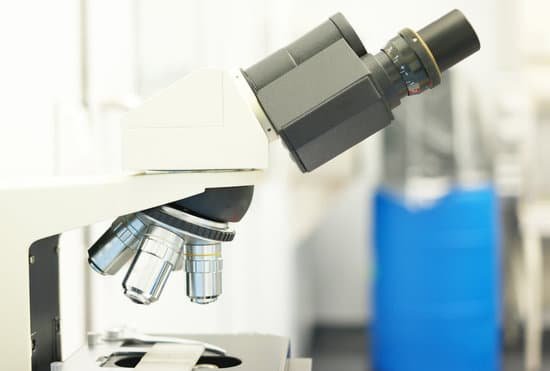
Why is everything backwards in a microscope? There are also mirrors in the microscope, which cause images to appear upside down and backwards. … The letter appears upside down and backwards because of two sets of mirrors in the microscope. This means that the slide must be moved in the opposite direction that you want the image to move.
What does inversion mean when using a microscope? Inversion is the reversal of an image projected by a microscope. Most microscopes used today are compound microscopes, meaning they have more that one lens involved in the magnification process. A light source underneath the sample projects light upward through the sample and into an objective lens.
What does it mean that the image is inverted when you look through the ocular lenses? What does it mean that the image is inverted when you look through the ocular lenses? The ocular lens or eyepiece lens acts as a magnifying glass for the image, the ocular lens makes the light rays spread more so that they appear to come from a larger inverted image beyond the objective lands.
What is the eyepiece of a microscope? The eyepiece, or ocular, magnifies the primary image produced by the objective; the eye can then use the full resolution capability of the objective. The microscope produces a virtual image of the specimen at the point of most distinct vision, generally 250 mm (10 in.) from the eye.
Why is everything backwards in a microscope? – Related Questions
What is the base used function in the microscope?
Base: The bottom of the microscope, used for support Illuminator: A steady light source (110 volts) used in place of a mirror.
Why do we use mineral oil for microscopes?
In light microscopy, oil immersion is a technique used to increase the resolving power of a microscope. This is achieved by immersing both the objective lens and the specimen in a transparent oil of high refractive index, thereby increasing the numerical aperture of the objective lens.
When to use stereoscopic microscope?
The stereo microscope is often used to study the surfaces of solid specimens or to carry out close work such as dissection, microsurgery, watch-making, circuit board manufacture or inspection, and fracture surfaces as in fractography and forensic engineering.
What medications can cause microscopic blood in the urine?
Drugs — Hematuria can be caused by medications, such as blood thinners, including heparin, warfarin (Coumadin) or aspirin-type medications, penicillins, sulfa-containing drugs and cyclophosphamide (Cytoxan).
Can you see dna with a compound microscope?
Given that DNA molecules are found inside the cells, they are too small to be seen with the naked eye. For this reason, a microscope is needed. While it is possible to see the nucleus (containing DNA) using a light microscope, DNA strands/threads can only be viewed using microscopes that allow for higher resolution.
Can i use a microscope with my glasses on?
Eyeglass wearers requiring simple lenses with a spherical power can use the microscope with or without their glasses, provided that the diopter setting of the focusing eyepiece is sufficient.
Is temperature microscopic or macroscopic concept?
Sol: Temperature is a macroscopic concept . This means that temperature is an average property of the large number of molecules which constitute a system . We can not define the temperature of a single molecule .
What is the price of compound microscope?
The most popular compound microscopes from some of the most well-known brands cost on average around $900-$1,200, although there are beginner microscopes that are just above the toy level that cost $100.
Why do i have microscopic hematuria?
The most common causes of microscopic hematuria are urinary tract infection, benign prostatic hyperplasia, and urinary calculi. However, up to 5% of patients with asymptomatic microscopic hematuria are found to have a urinary tract malignancy.
What is the purpose of staining a microscope specimen?
The most basic reason that cells are stained is to enhance visualization of the cell or certain cellular components under a microscope. Cells may also be stained to highlight metabolic processes or to differentiate between live and dead cells in a sample.
What is the magnification and resolution of a light microscope?
The maximum magnification of light microscopes is usually ×1500, and their maximum resolution is 200nm, due to the wavelength of light. An advantage of the light microscope is that it can be used to view a variety of samples, including whole living organisms or sections of larger plants and animals.
Why can t ribosomes be seen through a light microscope?
Some cell parts, including ribosomes, the endoplasmic reticulum, lysosomes, centrioles, and Golgi bodies, cannot be seen with light microscopes because these microscopes cannot achieve a magnification high enough to see these relatively tiny organelles.
Why was the compound microscope made?
A Dutch father-son team named Hans and Zacharias Janssen invented the first so-called compound microscope in the late 16th century when they discovered that, if they put a lens at the top and bottom of a tube and looked through it, objects on the other end became magnified. … “The hand lenses were much better.”
What is a polarizing light microscope forensics?
Polarized light microscopy (PLM) is a technique commonly used in the field of forensic science. PLM characterizes and identifies trace evidence found at crime scenes, such as fibers, hairs, paints, and glass fragments.
How to view sperm with a microscope?
You can view sperm at 400x magnification. You do NOT want a microscope that advertises anything above 1000x, it is just empty magnification and is unnecessary. In order to examine semen with the microscope you will need depression slides, cover slips, and a biological microscope.
What organelles can only be seen using an electron microscope?
Mitochondria are visible with the light microscope but can’t be seen in detail. Ribosomes are only visible with the electron microscope.
Who established the comparison microscope for use in firearms examination?
Phillip O. Gravelle developed the comparison microscope for use in firearm investigations with the assistance of Colonel Goddard in the early1920’s. An optical comparison microscope consists primarily of two relatively low powered, two-dimensional (2D) compound microscopes joined by an ocular unit or optical bridge.
Where are the lenses located in a compound microscope?
Typically, a compound microscope is used for viewing samples at high magnification (40 – 1000x), which is achieved by the combined effect of two sets of lenses: the ocular lens (in the eyepiece) and the objective lenses (close to the sample).
What is the medical definition of a microscope?
1 : an optical instrument consisting of a lens or combination of lenses for making enlarged images of minute objects especially : compound microscope — see light microscope, phase-contrast microscope, polarizing microscope, reflecting microscope, ultraviolet microscope.
How do you figure the total magnification of a microscope?
The total magnification of the microscope is calculated from the magnifying power of the objective multiplied by the magnification of the eyepiece and, where applicable, multiplied by intermediate magnifications. A distinction is made between magnification and lateral magnification.

