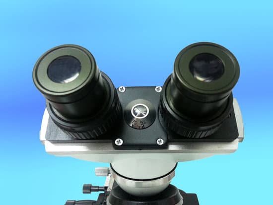
Why is staining useful when studying cells through a microscope? The most basic reason that cells are stained is to enhance visualization of the cell or certain cellular components under a microscope. Cells may also be stained to highlight metabolic processes or to differentiate between live and dead cells in a sample.
Why do we stain specimens before viewing them under a microscope? The main reason you stain a specimen before putting it under the microscope is to get a better look at it, but staining does much more than simply highlight the outlines of cells. Some stains can penetrate cell walls and highlight cell components, and this can help scientists visualize metabolic processes.
What is the importance of staining? Staining is used to highlight important features of the tissue as well as to enhance the tissue contrast. Hematoxylin is a basic dye that is commonly used in this process and stains the nuclei giving it a bluish color while eosin (another stain dye used in histology) stains the cell’s nucleus giving it a pinkish stain.
What are the advantages of staining the cheek cells? Having absorbed the stain, these parts of the cell become more visible under the microscope and can therefore be easily distinguished from other parts of the same cell. Without stains, cells would appear to be almost transparent, making it difficult to differentiate its parts.
Why is staining useful when studying cells through a microscope? – Related Questions
Which microscope should i buy?
You will need a compound microscope if you are viewing “smaller” specimens such as blood samples, bacteria, pond scum, water organisms, etc. … Typically, a compound microscope has 3-5 objective lenses that range from 4x-100x. Assuming 10x eyepieces and 100x objective, the total magnification would be 1,000 times.
What is a compound light microscope best for viewing?
Typically, a compound microscope is used for viewing samples at high magnification (40 – 1000x), which is achieved by the combined effect of two sets of lenses: the ocular lens (in the eyepiece) and the objective lenses (close to the sample).
Which microscope has the greatest working distance?
Low-power microscopes will have more generous stage working distances than high-power microscopes. Low-power microscopes will also be able to see more of the surface of the specimen, since the larger working distance and low power give them the ability to view more of the specimen at once.
How to identify e coli under a microscope?
When viewed under the microscope, Gram-negative E. Coli will appear pink in color. The absence of this (of purple color) is indicative of Gram-positive bacteria and the absence of Gram-negative E.
Which microscope used for histopathological image analysis?
The digital histopathological images are acquired through computerized electron microscope after tissue slide preparation. Different magnification images are used for different types of analysis; like for tissue classification low magnification (10X) and for cell segmentation and analysis higher magnification (40X).
What is the parts of microscope and their uses?
Tube: Connects the eyepiece to the objective lenses. Arm: Supports the tube and connects it to the base. Base: The bottom of the microscope, used for support. Illuminator: A steady light source (110 volts) used in place of a mirror.
Why ribosomes are not visible using a light microscope?
Ribosomes are not visible when using a light microscope because they are too small to see. They’re only about 25 nm while the maximum resolution of a light microscope is only 200 nm.
How did lister help develop the microscope?
J J Lister designed and made significantly improved microscope lenses free from achromatic aberration (where objects appear coloured) and spherical aberration (where all objects appear as if circular).
How is a light microscope work used and calculate magnification?
To calculate the total magnification of the compound light microscope multiply the magnification power of the ocular lens by the power of the objective lens. For instance, a 10x ocular and a 40x objective would have a 400x total magnification. The highest total magnification for a compound light microscope is 1000x.
What microscope would be used to look at swimming paramecium?
Using a student biological microscope (also known as a compound microscope), you can grow some paramecium and watch as they swim around just like the video below.
What is virtual image in microscope?
A simple microscope or magnifying glass (lens) produces an image of the object upon which the microscope or magnifying glass is focused. … Such images are termed virtual images and they appear upright, not inverted.
What does the diaphragm control on a microscope?
Opening and closing of the condenser aperture diaphragm controls the angle of the light cone reaching the specimen. The setting of the condenser’s aperture diaphragm, along with the aperture of the objective, determines the realized numerical aperture of the microscope system.
What is the function of mechanical stage in a microscope?
All microscopes are designed to include a stage where the specimen (usually mounted onto a glass slide) is placed for observation. Stages are often equipped with a mechanical device that holds the specimen slide in place and can smoothly translate the slide back and forth as well as from side to side.
Are there microscopic bugs in your eyebrows?
Speaking of mites that feed on human material, Demodex folliculorum (Simon) is one of three mite species living on your face. The microscopic critters are found across the human body, but are particularly dense near the nose, eyebrows and eyelashes.
Is everything under a microscope inverted?
Microscopes invert images which makes the picture appear to be upside down. The reason this happens is that microscopes use two lenses to help magnify the image.
Who invented digital microscope?
An early digital microscope was made by a lens company in Tokyo, Japan in 1986, which is now known as Hirox Co Ltd. It included a control box and a lens connected to a computer. Other versions of digital microscope were later developed by Keyence Corp and Leica Microsystems.
Do scanning electron microscopes have good resolution?
The apparatus has also a transmission operating mode (STEM mode). If the sample is thin enough, bright-field and dark-field images can be taken by the STEM detector, information content of which corresponds to those of TEM images. The ultimate resolution at this operating mode in ideal circumstances is 0.9 nm.
What is the significance of the microscope resolving power?
The resolving power of a microscope is the most important feature of the optical system and influences the ability to distinguish between fine details of a particular specimen.
What is the objective on a light microscope?
The objective lens of a microscope is the one at the bottom near the sample. At its simplest, it is a very high-powered magnifying glass, with very short focal length. This is brought very close to the specimen being examined so that the light from the specimen comes to a focus inside the microscope tube.
How to change objective lens on a microscope?
Turn the revolving turret (2) so that the lowest power objective lens (eg. 4x) is clicked into position. Place the microscope slide on the stage (6) and fasten it with the stage clips. Look at the objective lens (3) and the stage from the side and turn the focus knob (4) so the stage moves upward.
What are the illuminating parts of microscope and their functions?
In a modern microscope it consists of a light source, such as an electric lamp or a light-emitting diode, and a lens system forming the condenser. The condenser is placed below the stage and concentrates the light, providing bright, uniform illumination in the region of the object under observation.

