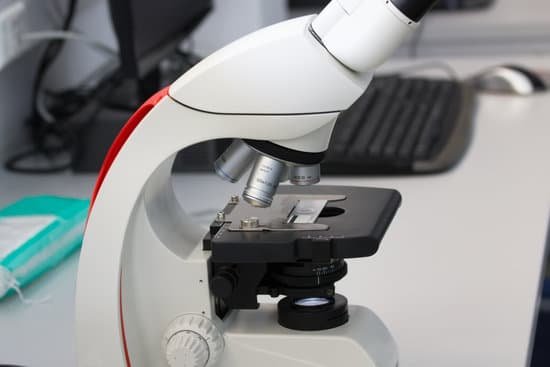
Why is the microscope an important tool for studying cells? Because most cells are too small to be seen by the naked eye, the study of cells has depended heavily on the use of microscopes. … Thus, the cell achieved its current recognition as the fundamental unit of all living organisms because of observations made with the light microscope.
What is the difference between upright and inverted microscope? An upright microscope focuses by moving the stage up and down. An inverted microscope has a fixed stage and the objectives move up and down to focus.
Why do we use inverted microscope? Inverted microscopes are useful for observing living cells or organisms at the bottom of a large container (e.g., a tissue culture flask) under more natural conditions than on a glass slide, as is the case with a conventional microscope.
What are the disadvantages of using an inverted microscope? The largest disadvantage is the cost. There are fewer manufacturers and the engineering to manufacture the microscope is more expensive. This means there are fewer used microscopes of this type on the market, and less competitive pricing.
Why is the microscope an important tool for studying cells? – Related Questions
Why does microscope invert the image?
Under the slide on which the object is being magnified, there is a light source that shines up and helps you to see the object better. This light is then refracted, or bent around the lens. Once it comes out of the other side, the two rays converge to make an enlarged and inverted image.
What is the resolution of a compound microscope?
The ocular lens is found in the eyepiece while the objective lens is found in the revolving nosepiece. The resolving power is the capacity of an instrument to resolve two points that are close together. The resolving power of a compound microscope is 0.25μm.
How small are the objects we see with a microscope?
The smallest thing that we can see with a ‘light’ microscope is about 500 nanometers. A nanometer is one-billionth (that’s 1,000,000,000th) of a meter. So the smallest thing that you can see with a light microscope is about 200 times smaller than the width of a hair.
How to increase resolution of microscope?
The resolution of a specimen viewed through a microscope can be increased by changing the objective lens. The objective lenses are the lenses that protrude downward over the specimen.
What does cardiac muscle tissue look like under a microscope?
Cardiac muscle tissue, like skeletal muscle tissue, looks striated or striped. The bundles are branched, like a tree, but connected at both ends. Unlike skeletal muscle tissue, the contraction of cardiac muscle tissue is usually not under conscious control, so it is called involuntary.
What cells can you see with a light microscope?
Using a light microscope, one can view cell walls, vacuoles, cytoplasm, chloroplasts, nucleus and cell membrane. Light microscopes use lenses and light to magnify cell parts.
Do air bubbles affect microscope?
The reasons why air bubbles can be problematic are: Bubbles hinder the free movement of organisms, such as ciliates. The bubbles cause optical artifacts at the place where the air meets the water. … The microscope optics are designed to give optimum resolution for a specimen which is surrounded by water.
How to identify bacteria in microscope?
Upon viewing the bacteria under the microscope, you will be able to identify the bacteria based on a wide variety of physical characteristics. This mainly involves looking at their shape and size. There are a wide variety of different shapes, yet the three main types are cocci, bacilli, and spiral.
What are the advantages of transmission electron microscope?
The advantage of the transmission electron microscope is that it magnifies specimens to a much higher degree than an optical microscope. Magnification of 10,000 times or more is possible, which allows scientists to see extremely small structures.
What can you see in an electron microscope?
Electron microscopes are used to investigate the ultrastructure of a wide range of biological and inorganic specimens including microorganisms, cells, large molecules, biopsy samples, metals, and crystals.
Why must cells stay microscopic in size?
Cells are microscopic, meaning they can’t be seen with the naked eye. … The reason cells can grow only to a certain size has to do with their surface area to volume ratio. Here, surface area is the area of the outside of the cell, called the plasma membrane. The volume is how much space is inside the cell.
How do electron microscopes produce an image?
The electron beam follows a vertical path through the microscope, which is held within a vacuum. … Detectors collect these X-rays, backscattered electrons, and secondary electrons and convert them into a signal that is sent to a screen similar to a television screen. This produces the final image.
What is the approximate minimum resolution of a light microscope?
The resolution of the light microscope cannot be small than the half of the wavelength of the visible light, which is 0.4-0.7 µm. When we can see green light (0.5 µm), the objects which are, at most, about 0.2 µm.
Why is visible light the limitation of compound microscope?
Since the microscope uses visible light and visible light has a set range of wavelengths. The microscope can’t produce the image of an object that is smaller than the length of the light wave. Any object that’s less than half the wavelength of the microscope’s illumination source is not visible under that microscope.
Can microscopic colitis get worse?
Microscopic colitis sometimes gets better on its own. If your symptoms continue without improvement or if they worsen, your doctor may recommend dietary changes before moving on to medications and other treatments.
How do microscopes work quizlet?
How do microscopes work? Use lenses to magnify the image of an object by focusing light or electrons. … It makes the image even larger.
What do yeast cells look like under a brightfield microscope?
When viewing the specimen under high magnification (1000x and above) one will see oval (egg shaped) organism, which are the yeast. It is also possible to observe the buds, which can be seen on some of the yeast cells.
How are electron beams focused in electron microscope?
The electron microscope uses a beam of electrons and their wave-like characteristics to magnify an object’s image, unlike the optical microscope that uses visible light to magnify images. … This beam is focused onto the sample using a magnetic lens.
How much do transmission electron microscopes cost?
The cost of a transmission electron microscope (TEM) can range from $300,000 to $10,000,000. The cost of a focused ion beam electron microscope (FIB) can range from $500,000 to $4,000,000. There can be a high degree of variation in the cost of an electron microscope between manufacturers and models.
Is the endoplasmic reticulum visible under a light microscope?
Some cell parts, including ribosomes, the endoplasmic reticulum, lysosomes, centrioles, and Golgi bodies, cannot be seen with light microscopes because these microscopes cannot achieve a magnification high enough to see these relatively tiny organelles.

