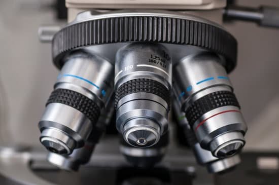
Why use a stereoscopic microscope over a compound? A compound microscope is generally used to view very small specimens or objects that you couldn’t normally see with the naked eye. A stereo microscope on the other hand is generally used to inspect larger objects such as small mechanical pieces, minerals, insects, and more.
Why would a researcher use a stereoscope over a compound light microscope? Here, stereo microscopes are used to view larger, opaque specimens while compound microscopes are used to view smaller, thin specimens. In the case of stereo microscopes, the specimen/object is too large to allow light to pass through. As such, they are particularly suitable for viewing surfaces of given objects.
When would you use a stereomicroscope instead of a compound microscope and why? A compound microscope is commonly used to view something in detail that you can’t see with the naked eye, such as bacteria or cells. A stereo microscope is typically used to inspect larger, opaque, and 3D objects, such as small electronic components or stamps.
When would you use a stereoscopic microscope? The stereo microscope is often used to study the surfaces of solid specimens or to carry out close work such as dissection, microsurgery, watch-making, circuit board manufacture or inspection, and fracture surfaces as in fractography and forensic engineering.
Why use a stereoscopic microscope over a compound? – Related Questions
How does a scanning electron microscope form a magnified image?
The SEM is an instrument that produces a largely magnified image by using electrons instead of light to form an image. A beam of electrons is produced at the top of the microscope by an electron gun. The electron beam follows a vertical path through the microscope, which is held within a vacuum.
What is the most commonly used microscope in the clinic?
One of the most common microscopes is the light microscope. These use light to illuminate an image, while one or sometimes multiple lenses magnify the specimen.
What does a fine focus do on a microscope do?
Focus (fine), Use the fine focus knob to sharpen the focus quality of the image after it has been brought into focus with the coarse focus knob. Illuminator, There is an illuminator built into the base of most microscopes. … Magnification, The degree to which the image of the specimen is enlarged by the objective.
How to use a virtual microscope?
When you open the microscope, go to the Catalogue and find the collection of slides that you need. The set of slides will be loaded onto the light-box. Passing the pointer over each thumbnail on the light box will pop-up a description of the slide. Click on the slide to place it onto the microscope stage.
How expensive are microscopes?
The most popular compound microscopes from some of the most well-known brands cost on average around $900-$1,200, although there are beginner microscopes that are just above the toy level that cost $100.
What was his microscope called?
Galileo Galilei soon improved upon the compound microscope design in 1609. Galileo called his device an occhiolino, or “little eye.”
How has microscopes helped us?
Microscopes allow humans to see cells that are too tiny to see with the naked eye. Therefore, once they were invented, a whole new microscopic world emerged for people to discover. On a microscopic level, new life forms were discovered and the germ theory of disease was born.
Why can you not see endoplasmic on a light microscope?
Some cell parts, including ribosomes, the endoplasmic reticulum, lysosomes, centrioles, and Golgi bodies, cannot be seen with light microscopes because these microscopes cannot achieve a magnification high enough to see these relatively tiny organelles.
How does microscope achieve magnification?
A microscope is an instrument that can be used to observe small objects, even cells. The image of an object is magnified through at least one lens in the microscope. This lens bends light toward the eye and makes an object appear larger than it actually is.
What 2 adjustments are on a standard microscope?
The coarse adjustment knob is the bigger of the two knobs and is located closest to the arm of the microscope. The fine adjustment knob is the smaller of the smaller of the two knobs and is located further away from the arm of the microscope.
How are light microscopes used?
A light microscope is a biology laboratory instrument or tool, that uses visible light to detect and magnify very small objects, and enlarging them. They use lenses to focus light on the specimen, magnifying it thus producing an image. … The image is then passed through one or two lenses for magnification for viewing.
What is a binocular dissecting microscope used for?
Stereomicroscope. A stereomicroscope, sometimes called a dissecting microscope or a binocular inspection microscope, is a low-power compound instrument used for a closer examination of three-dimensional specimens than is possible with a hand lens (Figure 1).
Why can t electron microscopes view living cells?
Electron microscopes are the most powerful type of microscope, capable of distinguishing even individual atoms. However, these microscopes cannot be used to image living cells because the electrons destroy the samples. … Damage would be avoided because the electrons would never actually hit the imaged objects.
How much can a confocal microscope magnify?
In general, the maximum magnification of a confocal microscope is 1000x, assuming the use of a combination of a 100x objective and a 10x ocular. It is dependent on the limitations of the mounting type used, thickness of tissue, and optics of the system.
How to collect microscopic sample from cow plant?
To create specific Microscope Prints from Samples you will need to gather slides from Plants, Fossils and Crystals. Choose the option ‘Collect Microscope Sample’ on the item. Level 2 Logic Skill will unlock Collect Microscope Samples from Plants. You can click on any full grown plant to collect microscope sample.
What is the magnification of a confocal microscope?
In general, the maximum magnification of a confocal microscope is 1000x, assuming the use of a combination of a 100x objective and a 10x ocular. It is dependent on the limitations of the mounting type used, thickness of tissue, and optics of the system.
How does a scanning electron microscope magnify an image?
The electron microscope uses a beam of electrons and their wave-like characteristics to magnify an object’s image, unlike the optical microscope that uses visible light to magnify images.
What is meant by resolving power of microscope?
Resolving power denotes the smallest detail that a microscope can resolve when imaging a specimen; it is a function of the design of the instrument and the properties of the light used in image formation. … The smaller the distance between the two points that can be distinguished, the higher the resolving power.
Can bacteria tell their under a microscope?
Bacteria are difficult to see with a bright-field compound microscope for several reasons: They are small: In order to see their shape, it is necessary to use a magnification of about 400x to 1000x.
How does the transmission microscope work?
How does TEM work? An electron source at the top of the microscope emits electrons that travel through a vacuum in the column of the microscope. Electromagnetic lenses are used to focus the electrons into a very thin beam and this is then directed through the specimen of interest.
What is a condenser lens on a microscope?
On upright microscopes, the condenser is located beneath the stage and serves to gather wavefronts from the microscope light source and concentrate them into a cone of light that illuminates the specimen with uniform intensity over the entire viewfield.

