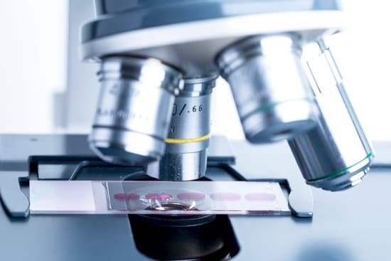
How does transmission electron microscope works? How does TEM work? An electron source at the top of the microscope emits electrons that travel through a vacuum in the column of the microscope. Electromagnetic lenses are used to focus the electrons into a very thin beam and this is then directed through the specimen of interest.
How is an image formed in a transmission electron microscope? Transmission electron microscopy (TEM) is a microscopy technique in which a beam of electrons is transmitted through a specimen to form an image. … An image is formed from the interaction of the electrons with the sample as the beam is transmitted through the specimen.
How does a doe TEM work? TEMs work by using a tungsten filament to produce an electron beam in a vacuum chamber. The emitted electrons are accelerated through an electromagnetic field that also narrowly focuses the beam. The beam is then passed through the sample material.
What is transmission electron microscope? Transmission electron microscopes (TEM) are microscopes that use a particle beam of electrons to visualize specimens and generate a highly-magnified image. TEMs can magnify objects up to 2 million times.
How does transmission electron microscope works? – Related Questions
What are microscopic algae called?
Microalgae or microphytes are microscopic algae invisible to the naked eye. They are phytoplankton typically found in freshwater and marine systems, living in both the water column and sediment. They are unicellular species which exist individually, or in chains or groups.
How to choose a compound microscope?
When buying a compound microscope, always ensure that the microscope has an iris diaphragm and good quality condenser – ideally, an Abbe condenser which allows for greater adjustments. Both items are found in the sub-stage of the microscope and are used in adjusting the base illumination.
What are the advantages and disadvantages of electron microscope?
Advantage: Light microscopes have high resolution. Electron microscopes are helpful in viewing surface details of a specimen. Disadvantage: Light microscopes can be used only in the presence of light and are costly. Electron microscopes uses short wavelength of electrons and hence have lower magnification.
How the microscope function?
A microscope is an instrument that can be used to observe small objects, even cells. The image of an object is magnified through at least one lens in the microscope. This lens bends light toward the eye and makes an object appear larger than it actually is.
What is a compound microscope used for in science?
Typically, a compound microscope is used for viewing samples at high magnification (40 – 1000x), which is achieved by the combined effect of two sets of lenses: the ocular lens (in the eyepiece) and the objective lenses (close to the sample).
What part of the atom is visible under the microscope?
The boundry where the electrons are located is the part of the atom that’s visible under the electron microscope.
How do electron microscopes help us understand cells?
The development of the electron microscopes therefore helped scientists to learn about the sub-cellular structures involved in aerobic respiration called mitochondria . The scientists developed their explanations about how the structure of the mitochondria allowed it to efficiently carry out aerobic respiration.
What does a chigger look like under a microscope?
Chiggers are barely visible to the naked eye (their length is less than 1/150th of an inch). A magnifying glass may be needed to see them. They are red in color and may be best appreciated when clustered in groups on the skin. The juvenile forms have six legs, although the (harmless) adult mites have eight legs.
Why is a microscope used in the staining technique?
The most basic reason that cells are stained is to enhance visualization of the cell or certain cellular components under a microscope. Cells may also be stained to highlight metabolic processes or to differentiate between live and dead cells in a sample.
What is the principle of travelling microscope?
A travelling microscope is an instrument for measuring length with a resolution typically in the order of 0.01mm. The precision is such that better-quality instruments have measuring scales made from Invar to avoid misreadings due to thermal effects.
What does microscopic mean in biology?
Microscopic. 1. Of extremely small size, visible only by the aid of the microscope. 2. Pertaining or relating to a microscope or to microscopy.
When using the microscope how is the focus adjusted?
So how does one focus a microscope? To focus a microscope, rotate to the lowest-power objective, and place your sample under the stage clips. Play with the magnification using the coarse adjustment knob and move your slide around until it is centered.
What is a direct microscopic count?
Direct Microscopic Count (DMC) is a quantitative test and is helpful in assessing the actual number of bacteria present in milk. … The method is useful for rapid estimation of the total bacterial population of a sample of milk and also in giving useful information for tracing the sources of contamination of milk.
How is a microscope properly transport?
When transporting the microscope, hold it in an upright position with one hand on its arm and the other supporting its base. Avoid jarring the instrument when setting it down. Use only special grit-free lens paper to clean the lenses.
Who made first compound microscope?
A Dutch father-son team named Hans and Zacharias Janssen invented the first so-called compound microscope in the late 16th century when they discovered that, if they put a lens at the top and bottom of a tube and looked through it, objects on the other end became magnified.
How to read microscope objective?
Microscope objective lenses will often have four numbers engraved on the barrel in a 2×2 array. The upper left number is the magnification factor of the objective. For example, 4x, 10x, 40x, and 100x. The upper right number is the numerical aperture of the objective.
Why are electron microscopes capable of revealing details?
As the wavelength of an electron can be up to 100,000 times shorter than that of visible light photons, electron microscopes have a higher resolving power than light microscopes and can reveal the structure of smaller objects.
Can stress cause onset of microscopic colitis?
This isn’t in your head. Stress is one of the factors that contribute to a colitis flare-up, along with tobacco smoking habits, diet, and your environment. Ulcerative colitis is an autoimmune disease that affects the large intestine (also known as your colon).
What happens when the objective of a microscope changes?
Changing from low power to high power increases the magnification of a specimen. The amount an image is magnified is equal to the magnification of the ocular lens, or eyepiece, multiplied by the magnification of the objective lens.
What is lens paper for a microscope?
Microscope Lens Paper is soft, dust-free paper that is used for cleaning microscope slides and lenses without scratching the glass.
How microscopes works?
A simple light microscope manipulates how light enters the eye using a convex lens, where both sides of the lens are curved outwards. When light reflects off of an object being viewed under the microscope and passes through the lens, it bends towards the eye. This makes the object look bigger than it actually is.

