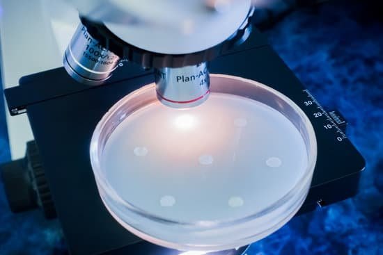
Can you use a handkerchief to clean a microscope? Use only lens paper or gauze and cleaning solution. Never use your finger, handkerchief, paper towels or spit to clean the lenses. Do not remove any parts for cleaning; it only allows dust to enter the microscope.
What are the materials that you can use to clean a microscope? Put a small amount of lens cleaning fluid or cleaning mixture on the tip of the lens paper. We recommend 70% ethanol because it can effectively and safely clean and disinfect the surface. Larger surfaces, such as a glass plate, may be too large to wipe using this technique.
What can be safely used to clean microscope lenses? Isopropyl alcohol is one of the best solvents but it must be at least 90%+ pure (do not use rubbing alcohol, 30% water). Everclear which is grain alcohol (you must be 21!) can also be used but it doesn’t do as well in dissolving crud.
Why oil is used in 100x microscope? The 100x lens is immersed in a drop of oil placed on the slide in order to eliminate any air gaps and lossof light due to refraction (bending of the light) as the light passes from glass (slide) → air → glass (objective lens). Immersion oil has the same refractive index of glass.
Can you use a handkerchief to clean a microscope? – Related Questions
How to see bacteria with a microscope?
Viewing bacteria under a microscope is much the same as looking at anything under a microscope. Prepare the sample of bacteria on a slide and place under the microscope on the stage. Adjust the focus then change the objective lens until the bacteria come into the field of view.
What microscope can see atoms?
An electron microscope can be used to magnify things over 500,000 times, enough to see lots of details inside cells. There are several types of electron microscope. A transmission electron microscope can be used to see nanoparticles and atoms.
What is direct microscopic count?
Direct Microscopic count (DMC) is a quantitative test and used to enumerate the number of bacterial clumps or somatic cells present in milk. This method is also used for the analysis of foods, water, and, in some cases, air for quantitative counting of microorganisms.
How does confocal microscope use laser light?
Similar to the widefield microscope, the confocal microscope uses fluorescence optics. Instead of illuminating the whole sample at once, laser light is focused onto a defined spot at a specific depth within the sample. … By scanning the specimen in a raster pattern, images of one single optical plane are created.
What is a turret on a microscope?
Turret or Objective Turret: The rotatable metal piece into which the microscope’s objective lenses are attached. A “turret” style stereo microscope refers to the type that has more than one objective lens which can then be rotated into position.
What are the strengths and weaknesses of a light microscope?
Advantage: Light microscopes have high magnification. Electron microscopes are helpful in viewing surface details of a specimen. Disadvantage: Light microscopes can be used only in the presence of light and have lower resolution. Electron microscopes can be used only for viewing ultra-thin specimens.
Does a light microscope produce a 3d image?
Stereo 3D microscopes produce real-time 3D images, but they are usually limited to low-magnification applications, such as dissection. Most compound light microscopes produce flat, 2D images because high-magnification microscope lenses have inherently shallow depth of field, rendering most of the image out of focus.
What does magnification mean on a microscope?
Magnification is the ability of a microscope to produce an image of an object at a scale larger (or even smaller) than its actual size.
How was the first compound microscope different from leeuwenhoeks microscope?
Whereas van Leeuwenhoek used a simple microscope, in which light is passed through just one lens, Galileo’s compound microscope was more sophisticated, passing light through two sets of lenses.
How to improve resolving power of microscope?
One way of increasing the optical resolving power of the microscope is to use immersion liquids between the front lens of the objective and the cover slip. Most objectives in the magnification range between 60x and 100x (and higher) are designed for use with immersion oil.
How to identify prophase under a microscope?
When you look at a cell in prophase under the microscope, you will see thick strands of DNA loose in the cell. If you are viewing early prophase, you might still see the intact nucleolus, which appears like a round, dark blob.
How to properly handle a light microscope?
When carrying the light microscope, handlers must put one hand on the base at all times, to avoid dropping it, while the other hand should be on the arm. The microscope must never be carried upside down, since the ocular will fall out. It should never be swung when it is carried, according to Miami University.
Can you see microbes without a microscope?
Yes. Most bacteria are too small to be seen without a microscope, but in 1999 scientists working off the coast of Namibia discovered a bacterium called Thiomargarita namibiensis (sulfur pearl of Namibia) whose individual cells can grow up to 0.75mm wide.
What shape are cardiac muscle cells microscope?
Basically, cardiac muscle cells are striated and arranged in cylindrical form with densely packed myofibrils over the length. Each cell is arranged to form a multiple connection with neighboring cells via intercalated discs (Fig.
What is a second type of scanning probe microscope?
There are several different types of scanning probe microscopes, the most prominent of which are atomic force microscopy (AFM) and scanning tunneling microscopy (STM). … Throughout the process, the microscope senses the distance of the probe tip from the surface of the sample.
How to put a slide on a microscope?
Turn the revolving turret (2) so that the lowest power objective lens (eg. 4x) is clicked into position. Place the microscope slide on the stage (6) and fasten it with the stage clips. Look at the objective lens (3) and the stage from the side and turn the focus knob (4) so the stage moves upward.
How to seal a microscope slide?
Place the coverslip with the sample pointing towards the slide onto the slide. Dip the brush of the nail polish into the bottle and then dry it a little bit by squeezing gently against the inner side of the bottle neck. Place four small dots of nail polish first on the corners of the coverslip to fix it a little bit.
Did robert hooke create a microscope?
Although Hooke did not make his own microscopes, he was heavily involved with the overall design and optical characteristics. The microscopes were actually made by London instrument maker Christopher Cock, who enjoyed a great deal of success due to the popularity of this microscope design and Hooke’s book.
When was the first electron microscope built?
Ernst Ruska at the University of Berlin, along with Max Knoll, combined these characteristics and built the first transmission electron microscope (TEM) in 1931, for which Ruska was awarded the Nobel Prize for Physics in 1986.
How would one improve the resolution of the microscope?
The resolution of a specimen viewed through a microscope can be increased by changing the objective lens. The objective lenses are the lenses that protrude downward over the specimen.

