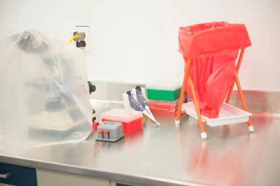
Does a stereo microscope invert the image? Microscopes invert images which makes the picture appear to be upside down. The reason this happens is that microscopes use two lenses to help magnify the image. Some microscopes have additional magnification settings which will turn the image right-side-up.
What does a stereo microscope observe? Stereo Microscopes enable 3D viewing of specimens visible to the naked eye. They are commonly known as Low Power or Dissecting Microscopes. An estimated 99% of stereo applications employ less than 50x magnification. Use them for viewing insects, crystals, plant life, circuit boards etc.
How do stereo microscopes form images? How Do Stereo Microscopes Work? A stereo or a dissecting microscope uses reflected light from the object. It magnifies at a low power hence ideal for amplifying opaque objects. Since it uses light that naturally reflects from the specimen, it is helpful to examine solid or thick samples.
Why are microscope images inverted? The ocular lens makes the light rays spread more, so that they appear to come from a large inverted image beyond the objective lens. Because light rays do not actually pass through this location, the image is called a virtual image.
Does a stereo microscope invert the image? – Related Questions
What microscope controls do you use to adjust field diaphragm?
AFTER THE SPECIMEN IS IN FOCUS AND THE CONDENSER PROPERLY ADJUSTED, IT IS TIME TO BRING THE FIELD DIAPHRAGM INTO FOCUS AND CENTER IT. 9. FIRST CLOSE THE FIELD DIAPHRAGM TO IT’S SMALLEST OPENING BY TURNING THE KNURLED ADJUSTMENT KNOB LOCATED BEHIND THE FIELD LENS ASSEMBLY IN THE BASE OF YOUR MICROSCOPE.
How do microscopes help us?
Microscopes are the tools that allow us to look more closely at objects, seeing beyond what is visible with the naked eye. Without them, we would have no idea about the existence of cells or how plants breathe or how rocks change over time.
What is a limitation of the light microscope?
The principal limitation of the light microscope is its resolving power. Using an objective of NA 1.4, and green light of wavelength 500 nm, the resolution limit is ∼0.2 μm. This value may be approximately halved, with some inconvenience, using ultraviolet radiation of shorter wavelengths.
What is magnification and resolution of a microscope?
Magnification is the ability to make small objects seem larger, such as making a microscopic organism visible. Resolution is the ability to distinguish two objects from each other. Light microscopy has limits to both its resolution and its magnification.
What is illuminator microscope?
Illuminator. There is an illuminator built into the base of most microscopes. The purpose of the illuminator is to provide even, high intensity light at the place of the field aperture, so that light can travel through the condensor to the specimen.
How can you calculate magnification on a microscope?
It’s very easy to figure out the magnification of your microscope. Simply multiply the magnification of the eyepiece by the magnification of the objective lens. The magnification of both microscope eyepieces and objectives is almost always engraved on the barrel (objective) or top (eyepiece).
Which type of microscope can produce three dimensional images?
The scanning electron microscope (SEM) lets us see the surface of three-dimensional objects in high resolution. It works by scanning the surface of an object with a focused beam of electrons and detecting electrons that are reflected from and knocked off the sample surface.
What type of microscope is used to visualize bacteria?
The compound microscope can be used to view a variety of samples, some of which include: blood cells, cheek cells, parasites, bacteria, algae, tissue, and thin sections of organs. Compound microscopes are used to view samples that can not be seen with the naked eye.
How to measure specimen under microscope?
The measurement of specimen size with a microscope is normally made by using an eyepiece graticule sometimes referred to as a reticule. This is a x10 eyepiece that has a scale inserted which is in focus at the same time as the specimen.
Can paramecium be seen with a light microscope?
Paramecium was probably one of the first single-celled organisms observed with a light microscope by the Dutch cloth vendor and amateur lens maker Antoni van Leuwenhoek (1632-1723) (Dobell, 1932), and it is still being investigated in the 21st century in the days of the modern electron microscopes.
Who developed a comparison microscope to compare bullets?
Philip O. Gravelle, a chemist, developed a comparison microscope for use in the identification of fired bullets and cartridge cases with the support and guidance of forensic ballistics pioneer Calvin Goddard. It was a significant advance in the science of firearms identification in forensic science.
What is the function of the microscope condenser?
On upright microscopes, the condenser is located beneath the stage and serves to gather wavefronts from the microscope light source and concentrate them into a cone of light that illuminates the specimen with uniform intensity over the entire viewfield.
Which microscope can provide a 3d image?
The scanning electron microscope (SEM) lets us see the surface of three-dimensional objects in high resolution. It works by scanning the surface of an object with a focused beam of electrons and detecting electrons that are reflected from and knocked off the sample surface.
What did rohrer and binnig call their new microscope?
With the introduction of these changes, Binnig and Rohrer turned the instrument into a microscope and called it Scanning Tunneling Microscope. It is not clear how exactly they came to the idea of transforming their tunneling testing instrument into an STM, displaying images of surfaces on the atomic scale.
Does an electron microscope use magnetic field?
Atomic-resolution electron microscopes utilize high-power magnetic lenses to produce magnified images of the atomic details of matter. Doing so involves placing samples inside the magnetic objective lens, where magnetic fields of up to a few tesla are always exerted.
Where are electron microscopes used?
Electron microscopy (EM) is a technique for obtaining high resolution images of biological and non-biological specimens. It is used in biomedical research to investigate the detailed structure of tissues, cells, organelles and macromolecular complexes.
What is a scanning electron microscope used for in forensics?
Electron microscopy (EM) has a wide variety of applications in forensic investigation. Numerous crime-scene micro-traces, including glass and paint fragments, tool marks, drugs, explosives and gunshot residue (GSR) can be visually and chemically analyzed with scanning electron microscopy (SEM).
What is a microelectrode microscope and what does it measure?
Microelectrode arrays captures the field potential or activity across an entire population of cells, with far greater data points per well, detecting activity patterns that would otherwise elude traditional assays such as patch clamp electrophysiology which probes a single cell such as a neuron.
Are images flipped in microscopes?
Microscopes invert images which makes the picture appear to be upside down. The reason this happens is that microscopes use two lenses to help magnify the image. Some microscopes have additional magnification settings which will turn the image right-side-up.
What is the arrangement of a two lens microscope?
A compound microscope is composed of two lenses: an objective and an eyepiece. The objective forms the first image, which is larger than the object. This first image is inside the focal length of the eyepiece and serves as the object for the eyepiece. The eyepiece forms final image that is further magnified.
Who are the pioneers in the development of microscope?
In the late 16th century several Dutch lens makers designed devices that magnified objects, but in 1609 Galileo Galilei perfected the first device known as a microscope. Dutch spectacle makers Zaccharias Janssen and Hans Lipperhey are noted as the first men to develop the concept of the compound microscope.

