Does a transmission electron microscope use z stacks? The most recent one applied to serial section transmission electron microscopy (ssTEM), uses elastic deformations in an optimized manner along the z-stack7.
What is unique about transmission electron microscope? The main difference is that TEMs use electrons rather than light in order to magnify images. … Electron microscopes, on the other hand, can produce much more highly magnified images because the beam of electrons has a smaller wavelength which creates images of higher resolution.
What type of samples can be used in a transmission electron microscope? by transmission electron microscope (TEM) it is critical to use TEM samples of ultimate quality (perfectly embedded, mechanically pretreated and ion milled ones).
How does a transmission electron microscope work? How does TEM work? An electron source at the top of the microscope emits electrons that travel through a vacuum in the column of the microscope. Electromagnetic lenses are used to focus the electrons into a very thin beam and this is then directed through the specimen of interest.
Does a transmission electron microscope use z stacks? – Related Questions
How are scanning tunneling microscopes used?
The scanning tunneling microscope (STM) works by scanning a very sharp metal wire tip over a surface. By bringing the tip very close to the surface, and by applying an electrical voltage to the tip or sample, we can image the surface at an extremely small scale – down to resolving individual atoms.
What conclusions can be drawn from microscopic hair analysis?
Three general conclusions can be reached as a result of microscopic hair analysis: exclusion, no conclusion, or association. Within the categories of exclusion and association, there are two subcategories (see Gaudette 1985).
Why should the stage of a microscope be kept dry?
Keep the stage dry at all times. Fluids can damage metal parts and also make it difficult to move a slide around on the stage.
How much does a traditional microscope cost?
The most popular compound microscopes from some of the most well-known brands cost on average around $900-$1,200, although there are beginner microscopes that are just above the toy level that cost $100.
What restricts the resolution of the light microscope?
The resolution of the light microscope cannot be small than the half of the wavelength of the visible light, which is 0.4-0.7 µm. When we can see green light (0.5 µm), the objects which are, at most, about 0.2 µm.
What is compound microscope wikipedia?
Compound microscopes have at least two lenses. In a compound microscope, the lens closer to the eye is called the eyepiece. The lens at the other end is called the objective. The lenses multiply up, so a 10x eyepiece and a 40x objective together give 400x magnification.
What is stereo zoom microscope?
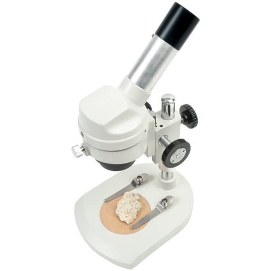
A stereo microscope is a type of optical microscope that allows the user to see a three-dimensional view of a specimen. Otherwise known as a dissecting microscope or stereo zoom microscope, the stereo microscope differs from the compound light microscope by having separate objective lenses and eyepieces.
How to see mites under microscope?
Place the slide glass side up on your microscope and look. The mites will be found at the end of the hair follicles. Use between 40x and 100x magnification. *Note: face mites desiccate quickly once removed from the face, within about five minutes, so look at the slide immediately.
What do air bubbles look like under a microscope?
The air bubbles possess a different refractive index than the surrounding medium (water). This makes the bubbles appear to have a thick dark border. The shape of the bubble focuses the light in such a way that the center of the bubble appears bright.
What is resolving power of electron microscope?
The geometrical aberrations of the objective lens of a scanning electron microscope limit the electron beam diameter to a few tens of angstroms. However, the resolving power generally obtained is of the order of 200–500 Å.
What can microscopic hematuria mean?
“Microscopic” means something is so small that it can only be seen through a special tool called a microscope. “Hematuria” means blood in the urine. So, if you have microscopic hematuria, you have red blood cells in your urine. These blood cells are so small, though, you can’t see the blood when you urinate.
Where is the biggest microscope located?
Lawrence Berkeley National Labs just turned on a $27 million electron microscope. Its ability to make images to a resolution of half the width of a hydrogen atom makes it the most powerful microscope in the world.
How to use an inverted microscope?
With an inverted microscope, you simply place your sample on the stage, focus onto the surface once and image it. Finished. The sample stays focused for all magnifications and further samples of the same sort are in focus alike.
What type of microscope is used to see bacteria?
The compound microscope can be used to view a variety of samples, some of which include: blood cells, cheek cells, parasites, bacteria, algae, tissue, and thin sections of organs. Compound microscopes are used to view samples that can not be seen with the naked eye.
What does microscopic mean dictionary?
1 : resembling a microscope especially in perception. 2a : invisible or indistinguishable without the use of a microscope. b : very small or fine or precise. 3 : of, relating to, or conducted with the microscope or microscopy.
How small does something have to be to be microscopic?
So, we can think of the microscopic scale as being from a millimetre (10-3 m) to a ten-millionth of a millimetre (10-10 m). Even within the microscopic scale, there are immense variations in the size of objects.
Can atoms be seen under an electron microscope?
Using electron microscopes, it is possible to image individual atoms. Summary: Scientists have calculated how it is possible to look inside the atom to image individual electron orbitals. An electron microscope can’t just snap a photo like a mobile phone camera can.
Where is the iris diaphragm located on a microscope?
Iris Diaphragm controls the amount of light reaching the specimen. It is located above the condenser and below the stage. Most high quality microscopes include an Abbe condenser with an iris diaphragm. Combined, they control both the focus and quantity of light applied to the specimen.
How many convex lenses are there in a compound microscope?
The simplest compound microscope is constructed from two convex lenses as shown schematically in Figure 2. The first lens is called the objective lens, and has typical magnification values from 5× to 100×.
Who observed cork cells under a microscope?
In 1665, Robert Hooke was the first to observe cork cells and their characteristic hexagonal shape, using the first optical microscope, which was invented by him at that time.
Does microscope show inverted image?
Microscopes invert images which makes the picture appear to be upside down. The reason this happens is that microscopes use two lenses to help magnify the image. Some microscopes have additional magnification settings which will turn the image right-side-up.

