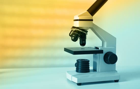
How did robert hooke invent the microscope? Hooke used his microscope to observe the smallest, previously hidden details of the natural world. His book Micrographia revealed and described his discoveries. … Hooke looked at the bark of a cork tree and observed its microscopic structure. In doing so, he discovered and named the cell – the building block of life.
How did Hooke invent the microscope? To combat dark specimen images, Hooke designed an ingenious method of concentrating light on his specimens, as shown in the illustration. He passed light generated from an oil lamp through a water-filled glass flask to diffuse the light and provide a more even and intense illumination for the samples.
How did Robert Hooke come up with his discovery? When he looked at a sliver of cork through his microscope, he noticed some “pores” or “cells” in it. Hooke believed the cells had served as containers for the “noble juices” or “fibrous threads” of the once-living cork tree.
How did Hooke’s compound microscope work? Hooke’s microscope was a much larger, ‘compound’ instrument. … A drawing of Hooke’s microscope, from Micrographia (1665). He used a glass globe filled with water to focus light from a small flame onto the specimen, to counteract the darkened images caused by lens aberrations. A replica of Leeuwenhoek’s simple microscope.
How did robert hooke invent the microscope? – Related Questions
What is the focal plane of a microscope?
A plane drawn perpendicular to the lens axis at the focal point is the focal plane. … The front focal plane of the eyepiece is the side inside the microscope. Back or Image Side The side of a lens where an image is formed is called the image side or back side of the lens.
How tell cells different animals apart microscope?
Under a microscope, plant cells from the same source will have a uniform size and shape. Beneath a plant cell’s cell wall is a cell membrane. An animal cell also contains a cell membrane to keep all the organelles and cytoplasm contained, but it lacks a cell wall.
Are roundworm eggs microscope?
Microscopes are not always available in poor communities, however, since they are difficult to transport and break easily. … The improvised microscope detected giant roundworm eggs 81 percent of the time, roundworm eggs 54 percent of the time and hookworm eggs 14 percent of the time.
How could we build a microscope with a higher resolution?
To achieve the maximum (theoretical) resolution in a microscope system, each of the optical components should be of the highest NA available (taking into consideration the angular aperture). In addition, using a shorter wavelength of light to view the specimen will increase the resolution.
How to look at mold under a microscope?
Place a drop of water in the center of the slide, using an eyedropper if you have one, or the tip of a clean finger. You can use solution of methylene blue instead, which is a microscope stain, and makes the sample easier to see by coloring certain parts of the mold cells.
How to use microscope sims 4?
Choose the option ‘Collect Microscope Sample’ on the item. Level 2 Logic Skill will unlock Collect Microscope Samples from Plants. You can click on any full grown plant to collect microscope sample. Level 5 Logic Skill will unlock Collect Microscope Samples from Fossils.
How big is hair under a microscope?
How big does hair look under the microscope? The answer varies per species, with the width of human hair averaging at around 70 micrometers.
How to adjust a microscope condenser?
Most compound light microscopes have a small knob (2) to raise and lower the condenser holder. Lower this holder so the condenser can slide into the holder below the stage. Once you have inserted the condenser, tighten the set screw (3) to hold the condenser in place.
Do smooth muscles appear striated under a microscope?
Skeletal muscles are long and cylindrical in appearance; when viewed under a microscope, skeletal muscle tissue has a striped or striated appearance. … Smooth muscle has no striations, is not under voluntary control, has only one nucleus per cell, is tapered at both ends, and is called involuntary muscle.
Who invented early microscopes?
Lens Crafters Circa 1590: Invention of the Microscope. Every major field of science has benefited from the use of some form of microscope, an invention that dates back to the late 16th century and a modest Dutch eyeglass maker named Zacharias Janssen.
Does a tem microscope study surface features?
The TEM is analogous in many ways to the conventional (compound) light microscope. … It provides detailed images of the surfaces of cells and whole organisms that are not possible by TEM. It can also be used for particle counting and size determination, and for process control.
What type of microscope is used to view muscle cells?
Compound microscopes are light illuminated. The image seen with this type of microscope is two dimensional. This microscope is the most commonly used. You can view individual cells, even living ones.
Can see a cell with a compound light microscope?
Explanation: The maximum magnification of a light compound microscope is 2000x. You can expect to see the cell nucleus and nucleolus, cytoplasm, cell membrane, cell walls and chloroplasts.
How many lenses does a microscope have?
A typical microscope has three or four objective lenses with different magnifications, screwed into a circular “nosepiece” which may be rotated to select the required lens.
What is the use of scanner in microscope?
A scanning electron microscope (SEM) scans a focused electron beam over a surface to create an image. The electrons in the beam interact with the sample, producing various signals that can be used to obtain information about the surface topography and composition.
What is used for first focusing on a microscope?
5. FOCUS ON SPECIMEN, FIRST USING THE COARSE AND THEN THE FINE FOCUS CONTROLS. YOU MAY HAVE TO MOVE THE SLIDE AROUND ON THE STAGE OF THE MICROSCOPE TO BRING THE SPECIMEN INTO THE VIEWING AREA.
What is the use of stage in microscope?
All microscopes are designed to include a stage where the specimen (usually mounted onto a glass slide) is placed for observation. Stages are often equipped with a mechanical device that holds the specimen slide in place and can smoothly translate the slide back and forth as well as from side to side.
Are microscope slides borosilicate or soda lime?
Microscope slides have to be made of optical quality glass. Some of the best microscope slides are made of soda-lime glass or borosilicate glass. Sometimes special optical quality transparent plastics are used.
What does microscope slide mean in biology?
A microscope slide is a thin flat piece of glass, typically 75 by 26 mm (3 by 1 inches) and about 1 mm thick, used to hold objects for examination under a microscope. Typically the object is mounted (secured) on the slide, and then both are inserted together in the microscope for viewing.
How to set up microscope condenser?
Most compound light microscopes have a small knob (2) to raise and lower the condenser holder. Lower this holder so the condenser can slide into the holder below the stage. Once you have inserted the condenser, tighten the set screw (3) to hold the condenser in place.
When focusing a microscope should begin with?
When focusing on a slide, ALWAYS start with either the 4X or 10X objective. Once you have the object in focus, then switch to the next higher power objective. Re-focus on the image and then switch to the next highest power.

