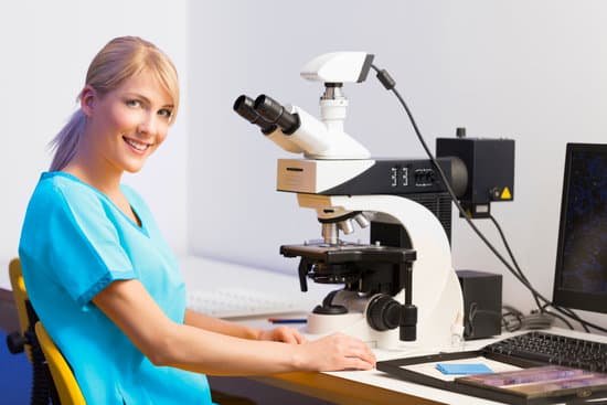
How does microscope help us? Microscopes are the tools that allow us to look more closely at objects, seeing beyond what is visible with the naked eye. Without them, we would have no idea about the existence of cells or how plants breathe or how rocks change over time.
What have microscopes helped us discover? Microscopes allow humans to see cells that are too tiny to see with the naked eye. Therefore, once they were invented, a whole new microscopic world emerged for people to discover. On a microscopic level, new life forms were discovered and the germ theory of disease was born.
What are the uses of microscope in your daily life? It is an instrument that magnifies objects in size so as to enable the naked eye to see things clearly. 2. They are helpful in creating electrician circuits due to their higher magnification abilities and help in creation of other electronic devices.
What are the 5 uses of microscope? The microscope is one of the most important tools used in chemistry and biology. This instrument allows a scientist or doctor to magnify an object to look at it in detail. Many types of microscopes exist, allowing different levels of magnification and producing different types of images.
How does microscope help us? – Related Questions
What is the aperture on a microscope?
Numerical Aperture and Resolution. The numerical aperture of a microscope objective is the measure of its ability to gather light and to resolve fine specimen detail while working at a fixed object (or specimen) distance. … The smaller the object, the more pronounced the diffraction of incident light rays will be.
What is coarse focus in microscope?
Focus (coarse), The coarse focus knob is used to bring the specimen into approximate or near focus. Focus (fine), Use the fine focus knob to sharpen the focus quality of the image after it has been brought into focus with the coarse focus knob.
What does calibrating the microscope mean?
Using a microscope that’s calibrated means that the same results will be produced on the exact same sample under the same conditions if you were to use an entirely different microscope that was also calibrated. The reason to calibrate is to get the most accurate measurement of your sample.
What does immersion oil do for a microscope?
Immersion Oil contributes to two characteristics of the image viewed through the microscope: finer resolution and brightness. These characteristics are most critical under high magnification; so it is only the higher power, short focus, objectives that are usually designed for oil immersion.
How to handle a light microscope properly?
When carrying the light microscope, handlers must put one hand on the base at all times, to avoid dropping it, while the other hand should be on the arm. The microscope must never be carried upside down, since the ocular will fall out. It should never be swung when it is carried, according to Miami University.
What microscope did leeuwenhoek use?
Antonie van Leeuwenhoek used single-lens microscopes, which he made, to make the first observations of bacteria and protozoa.
What can be viewed with transmitted light on a microscope?
Transmission light microscopes are used to look at thin sections – the specimen must transmit the light which includes tissues, cell walls, crystalline components, and thin films, etc.
What type of microscope has the highest magnification?
Out of all types of microscopes, the electron microscope has the greatest capability in achieving high magnification and resolution levels, enabling us to look at things right down to each individual atom.
Is staining required for a light microscope?
Biological samples for the light microscope (particularly compound microscopes) often need to be ‘stained’ (coloured) in some way to make it easier for users to understand what they’re seeing. This is important because these samples often lack contrast, which makes it hard to distinguish between parts of the sample.
Can a microscope see an atom?
Atoms are really small. So small, in fact, that it’s impossible to see one with the naked eye, even with the most powerful of microscopes. … Now, a photograph shows a single atom floating in an electric field, and it’s large enough to see without any kind of microscope.
Are c elegans microscope?
C. elegans allows to perform experiments involving large numbers of isogenic animals ensuring statistical robustness and making it a powerful model organism for genetic high-throughput screens. Additionally, it is one of the few model organisms that can be imaged in its entirety using electron microscopy (see Hall D.
How to use monocular microscope?
The proper way to use a monocular microscope is to look through the eyepiece with one eye and keep the other eye open (this helps avoid eye strain). Remember, everything is upside down and backwards. When you move the slide to the right, the image goes to the left!
How many objective lenses are on most student compound microscopes?
Objective Lenses: Usually you will find 3 or 4 objective lenses on a microscope. They almost always consist of 4x, 10x, 40x and 100x powers.
What is the difference between gross anatomy and microscopic anatomy?
“Gross anatomy” customarily refers to the study of those body structures large enough to be examined without the help of magnifying devices, while microscopic anatomy is concerned with the study of structural units small enough to be seen only with a light microscope. Dissection is basic to all anatomical research.
What is the arm of a microscope used for?
Arm connects to the base and supports the microscope head. It is also used to carry the microscope.
Can naproxen cause microscopic blood in urine?
If you can see blood in your urine, rest and don’t do any strenuous activity until your next exam. Don’t use aspirin, blood thinners, or anti-platelet or anti-inflammatory medicines. These include ibuprofen and naproxen. These thin the blood and may increase bleeding.
What year did the first microscope invented?
Lens Crafters Circa 1590: Invention of the Microscope. Every major field of science has benefited from the use of some form of microscope, an invention that dates back to the late 16th century and a modest Dutch eyeglass maker named Zacharias Janssen.
What is the microscopic structure of skeletal muscle tissue?
Skeletal muscle fibers are long, multinucleated cells. The membrane of the cell is the sarcolemma; the cytoplasm of the cell is the sarcoplasm. The sarcoplasmic reticulum (SR) is a form of endoplasmic reticulum. Muscle fibers are composed of myofibrils which are composed of sarcomeres linked in series.
What is 40x on microscope?
The total magnification of a high-power objective lens combined with a 10x eyepiece is equal to 400x magnification, giving you a very detailed picture of the specimen in your slide.
How to view dust mites under a microscope?
Dust mites are too small to see with the human eye, but can be seen at 20 times magnification with a microscope. You can easily calculate the total magnification of your microscope to see if it is strong enough to see the mite by multiplying the eyepiece magnification by the objective lens modification.
Do hematologists use microscopes?
Hematologists (alternate spelling “haematologists”) routinely investigate peripheral blood smears on glass slides with a microscope to find any abnormalities indicating hematological diseases or to look for blood parasites, such as those found for malaria and filariasis.

