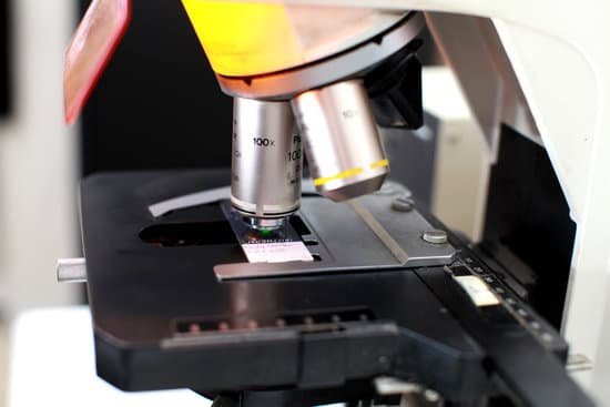
How microscope lenses work? A simple light microscope manipulates how light enters the eye using a convex lens, where both sides of the lens are curved outwards. When light reflects off of an object being viewed under the microscope and passes through the lens, it bends towards the eye. This makes the object look bigger than it actually is.
How does a basic 2 lens microscope work? A compound microscope uses two or more lenses to produce a magnified image of an object, known as a specimen, placed on a slide (a piece of glass) at the base. … By raising and lowering the stage, you move the lenses closer to or further away from the object you’re examining, adjusting the focus of the image you see.
What is the purpose of the lenses on a microscope? In microscopy, the objective lenses are the optical elements closest to the specimen. The objective lens gathers light from the specimen, which is focused to produce the real image that is seen on the ocular lens. Objective lenses are the most complex part of the microscope due to their multi-element design.
What are the 3 different lenses on a microscope? A typical compound microscope will have four objective lenses: one scanning lens, low-power lens, high-power lens, and an oil-immersion lens.
How microscope lenses work? – Related Questions
Can you see protein in a microscope?
In a conventional optical microscope, objects less than about 200 nanometers apart cannot be distinguished from one another. … Although electron microscopes produce a detailed image of very small structures, they cannot provide an image of the proteins that make up those structures.
What to do if cant find specimen on microscope?
If you cannot see anything, move the slide slightly while viewing and focusing. If nothing appears, reduce the light and repeat step 4. Once in focus on low power, center the object of interest by moving the slide. Rotate the objective to the medium power and adjust the fine focus only.
How to find working distance microscope?
There are two ways I would recommend finding the working distance of an objective lens. The first is to check to see if it is inscribed on the objective barrel. The inscription will be have the letters “WD” which is short for “working distance” followed by the length in millimeters like this: “WD: 0.5” or “0.5 EL WD”.
What to use a microscope for?
A microscope is an instrument that can be used to observe small objects, even cells. The image of an object is magnified through at least one lens in the microscope. This lens bends light toward the eye and makes an object appear larger than it actually is.
When did robert hooke discovered the microscope?
The discovery of the cell would not have been possible if not for advancements to the microscope. Interested in learning more about the microscopic world, scientist Robert Hooke improved the design of the existing compound microscope in 1665.
What is the field number of a microscope?
The field number (FN) in microscopy is defined as the diameter of the area in the intermediate image plane that can be observed through the eyepiece. A field number of, e.g., 20 mm indicates that the observed sample area after magnification by the objective lens is restricted to a diameter of 20 mm.
What part of the microscope helps adjust brightness?
Diaphragm or Iris: The diaphragm or iris is located under the stage and is an apparatus that can be adjusted to vary the intensity, and size, of the cone of light that is projected through the slide.
How have light microscopes changed over time?
The optical quality of lenses increased and the microscopes are similar to the ones we use today. Throughout their development, the magnification of light microscopes has increased, but very high magnifications are not possible. The maximum magnification with a light microscope is around ×1500.
What does phase contrast microscope do?
Phase contrast is a light microscopy technique used to enhance the contrast of images of transparent and colourless specimens. It enables visualisation of cells and cell components that would be difficult to see using an ordinary light microscope.
What is the magnification of the electron microscope?
This makes electron microscopes more powerful than light microscopes. A light microscope can magnify things up to 2000x, but an electron microscope can magnify between 1 and 50 million times depending on which type you use!
How does an electron microscope work simple?
The electron microscope uses a beam of electrons and their wave-like characteristics to magnify an object’s image, unlike the optical microscope that uses visible light to magnify images. … This stream is confined and focused using metal apertures and magnetic lenses into a thin, focused, monochromatic beam.
What does a polarizing light microscope do?
The polarized light microscope is designed to observe and photograph specimens that are visible primarily due to their optically anisotropic character.
What does it mean when a lab microscope is parfocal?
Parfocal means that the microscope is binocular. … Parfocal means that when one objective lens is in focus, then the other objectives will also be in focus.
What is the function of substage in compound microscope?
The substage condenser gathers light from the microscope light source and concentrates it into a cone of light that illuminates the specimen with uniform intensity over the entire viewfield.
What is the smallest object seen through a microscope?
The smallest thing that we can see with a ‘light’ microscope is about 500 nanometers. A nanometer is one-billionth (that’s 1,000,000,000th) of a meter. So the smallest thing that you can see with a light microscope is about 200 times smaller than the width of a hair. Bacteria are about 1000 nanometers in size.
Can microscopes see dna?
Given that DNA molecules are found inside the cells, they are too small to be seen with the naked eye. For this reason, a microscope is needed. While it is possible to see the nucleus (containing DNA) using a light microscope, DNA strands/threads can only be viewed using microscopes that allow for higher resolution.
How the first microscope was developed?
A Dutch father-son team named Hans and Zacharias Janssen invented the first so-called compound microscope in the late 16th century when they discovered that, if they put a lens at the top and bottom of a tube and looked through it, objects on the other end became magnified. … “The hand lenses were much better.”
How much does a tem microscope cost?
The cost of a transmission electron microscope (TEM) can range from $300,000 to $10,000,000. The cost of a focused ion beam electron microscope (FIB) can range from $500,000 to $4,000,000. There can be a high degree of variation in the cost of an electron microscope between manufacturers and models.
What do crabs look like under a microscope?
Under the microscope, pubic lice look like tiny crabs. To the naked eye, they appear to be pale gray, but get darker when swollen with blood. They attach themselves and their eggs to pubic hair, underarm hair, eyelashes, and eyebrows.
How to calculate low power magnification of a microscope?
To calculate the total magnification of the compound light microscope multiply the magnification power of the ocular lens by the power of the objective lens. For instance, a 10x ocular and a 40x objective would have a 400x total magnification.
What are types of microscopic colitis?
Two types of microscopic colitis are lymphocytic colitis and collagenous colitis. The two types cause different changes in colon tissue. In lymphocytic colitis, the colon lining contains more white blood cells than normal.

