How small is microscopic? So, we can think of the microscopic scale as being from a millimetre (10-3 m) to a ten-millionth of a millimetre (10-10 m). Even within the microscopic scale, there are immense variations in the size of objects.
Is microscopic smaller than tiny? is that tiny is very small while microscopic is of, or relating to microscopes or microscopy; microscopal.
What is the smallest microscopic item? Answer 1: The smallest object that we can see using a microscope (in a general sense) is atom, whose size is around 0.1 nano meter.
Is microscopic or macroscopic smaller? Unsourced material may be challenged and removed. The macroscopic scale is the length scale on which objects or phenomena are large enough to be visible with the naked eye, without magnifying optical instruments. It is the opposite of microscopic.
How small is microscopic? – Related Questions
What did robert hooke first look at under the microscope?
Hooke discovered the first known microorganisms, in the form of microscopic fungi, in 1665. This preceded Antonie van Leeuwenhoek’s discovery of single-celled life by nine years. Hooke looked at the bark of a cork tree and observed its microscopic structure.
Who made microscope first?
The development of the microscope allowed scientists to make new insights into the body and disease. It’s not clear who invented the first microscope, but the Dutch spectacle maker Zacharias Janssen (b. 1585) is credited with making one of the earliest compound microscopes (ones that used two lenses) around 1600.
What does the word compound microscope mean in science?
A compound microscope is a microscope that uses multiple lenses to enlarge the image of a sample. … The total magnification is calculated by multiplying the magnification of the ocular lens by the magnification of the objective lens. Light is passed through the sample (called transmitted light illumination).
How many types of lenses do compound light microscopes have?
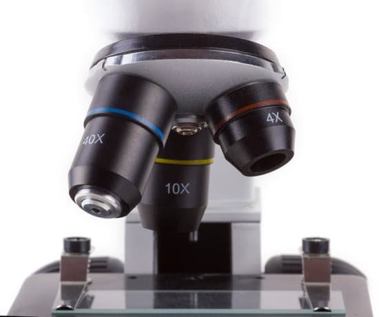
The lens that a person looks into is called the ocular lens and the lens nearest the specimen (pictured) is called the objective lens.
How to calculate resolving power of a microscope?
λ/2 NA), the specimen must be viewed using either shorter wavelength (λ) light or through an imaging medium with a relatively high refractive index or with optical components which have a high NA (or, indeed, a combination of all of these factors).
What are the microscopic air sacs in the lungs?
In your lungs, the main airways (bronchi) branch off into smaller and smaller passageways — the smallest, called bronchioles, lead to tiny air sacs (alveoli).
What are the parts of microscope and function?
Eyepiece Lens: the lens at the top that you look through, usually 10x or 15x power. Tube: Connects the eyepiece to the objective lenses. Arm: Supports the tube and connects it to the base. Base: The bottom of the microscope, used for support.
What does staph look like under microscope?
Staphylococcus aureus bacteria are pathogens to both man and other mammals. They are gram positive bacteria that are small round in shape (cocci) and occur as clusters appearing like a bunch of grapes on electron microscopy.
When carrying the microscope you should hold it by?
When carrying the microscope, hold its arm securely with both hands. When carrying the microscope, do not hold the focus knobs, eyepiece tube, stage, or other components as it may result in those parts coming off and cause of trouble.
How do i view my usb microscope?
To view the image from the USB microscope you will need to install webcam viewing software such as VLC media player (available for free from https://www.videolan.org/index.en-GB.html). Take the long metal rod with a thread on one end and start to thread it through the hole in the black base.
What two parts should you carry the microscope by?
When carrying a compound microscope always take care to lift it by both the arm and base, simultaneously. There are two optical systems in a compound microscope: Eyepiece Lenses and Objective Lenses: Eyepiece or Ocular is what you look through at the top of the microscope.
How have electron microscopes changed cell biology?
Electron microscopes provide higher magnification, higher resolution, and more detail than light microscopes. The unified cell theory states that one or more cells comprise all organisms, the cell is the basic unit of life, and new cells arise from existing cells.
How much magnification can usb microscopes?
The Plugable USB 2.0 Digital Microscope is one of the best USB microscopes with a 40 to 250x magnification power.
What kind of lens is used in compound microscope?
A compound microscope is made of two convex lenses; the first, the ocular lens, is close to the eye, and the second is the objective lens.
What is pseudopodia under a microscope?
Essentially, Pseudopodia are temporary projections of the cytoplasm that make it possible for amoebae to move. Pseudopods are some of the most distinguishable features of amoebae and their formation is based on the flow of the protoplasm.
What is simple microscope definition?
A simple microscope is a magnifying glass that has a double convex lens with a short focal length. The examples of this kind of instrument include the hand lens and reading lens. When an object is kept near the lens, then its principal focus with an image is produced, which is erect and bigger than the original object.
Why can you see dna under a microscope?
Given that DNA molecules are found inside the cells, they are too small to be seen with the naked eye. For this reason, a microscope is needed. While it is possible to see the nucleus (containing DNA) using a light microscope, DNA strands/threads can only be viewed using microscopes that allow for higher resolution.
How much does the eyepiece of a microscope magnify?
The eyepiece lens usually magnifies 10x, and a typical objective lens magnifies 40x. (Microscopes usually come with a set of objective lenses that can be interchanged to vary the magnification.)
How are electron microscopes used in workplaces?
Electron microscopy is often used for industrial purposes to assist in developing new products and throughout the manufacturing process. For example, electronics industries use electron microscopes for high-resolution imaging in the development and manufacturing processes of semiconductors and other electronics.
How do you determine the total magnification of a microscope?
The total magnification of the microscope is calculated from the magnifying power of the objective multiplied by the magnification of the eyepiece and, where applicable, multiplied by intermediate magnifications. A distinction is made between magnification and lateral magnification.
Which microscope lets you see at the highest resolution?
The microscope that can achieve the highest magnification and greatest resolution is the electron microscope, which is an optical instrument that is designed to enable us to see microscopic details down to the atomic scale (check also atom microscopy).

