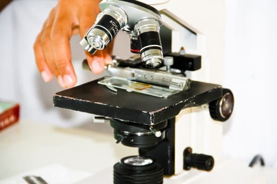
How to determine field of view for a microscope? For instance, if your eyepiece reads 10X/22, and the magnification of your objective lens is 40. First, multiply 10 and 40 to get 400. Then divide 22 by 400 to get a FOV diameter of 0.055 millimeters.
What is the field of view of a microscope? Introduction. Microscope field of view (FOV) is the maximum area visible when looking through the microscope eyepiece (eyepiece FOV) or scientific camera (camera FOV), usually quoted as a diameter measurement (Figure 1).
What is the field of view of a microscope at 40x? Field of view is how much of your specimen or object you will be able to see through the microscope. At 40x magnification you will be able to see 5mm. At 100x magnification you will be able to see 2mm. At 400x magnification you will be able to see 0.45mm, or 450 microns.
How do you find the area of the field of view? If the angle of the field of view is a degrees than you can see a/360 of the circle so the area of the sector you can view is (a/360) × (π r2) square units.
How to determine field of view for a microscope? – Related Questions
What part of a microscope focuses an image?
Condenser Lens: The purpose of the condenser lens is to focus the light onto the specimen. Condenser lenses are most useful at the highest powers (400x and above). Microscopes with in-stage condenser lenses render a sharper image than those with no lens (at 400x).
What you see through a microscope?
A microscope lets you look at and study very tiny things in great detail, which the naked eye cannot see. Even under a low-power optical microscope, the fine structures of specimens, or the objects under view, can be seen. … A photograph of the magnified view through a microscope is called a micrograph.
What is macroscopic and microscopic property?
The key difference between macroscopic and microscopic properties is that macroscopic properties are the properties of matter in bulk whereas microscopic properties are properties of the constituents of matter in bulk. The term microscopic refers to anything that is invisible to the naked eye.
How can resolving power of a microscope be improved?
One way of increasing the optical resolving power of the microscope is to use immersion liquids between the front lens of the objective and the cover slip. Most objectives in the magnification range between 60x and 100x (and higher) are designed for use with immersion oil.
How many lenses are in a microscope?
A typical microscope has three or four objective lenses with different magnifications, screwed into a circular “nosepiece” which may be rotated to select the required lens.
What is body tube in microscope?
The microscope body tube separates the objective and the eyepiece and assures continuous alignment of the optics. It is a standardized length, anthropometrically related to the distance between the height of a bench or tabletop (on which the microscope stands) and the position of the seated observer’s…
How to use vernier scale microscope?
To take a reading, first note where the ‘0’ on the Vernier scale meets the main scale. If it is positioned between numbers, the lower number should be used. Next, note where a mark on the smaller Vernier scale aligns directly with a mark on the main scale. This gives the decimal place figure (i.e 0.0 to 0.9).
Why do scientists use microscopes to study cells?
A cell is the smallest unit of life. Most cells are so small that they cannot be viewed with the naked eye. Therefore, scientists must use microscopes to study cells. Electron microscopes provide higher magnification, higher resolution, and more detail than light microscopes.
How much magnification to see stuff in microscope?
The compound microscope typically has three or four magnifications – 40x, 100x, 400x, and sometimes 1000x. At 40x magnification you will be able to see 5mm. At 100x magnification you will be able to see 2mm. At 400x magnification you will be able to see 0.45mm, or 450 microns.
What does the 40 on this microscope objective lens mean?
Coverslips with a deviating thickness will result is an image of lower resolution. 4, 10, 20, 40, 100: This represents the magnification of the objective. The total magnification is calculated by multiplying the magnification of the objective with the magnification of the ocular (eye piece), which is usually 10x.
Is bacteria multiple celled microscopic organisms?
Bacteria are multiple-celled microscopic organisms. Bacteria are the most abundant form of life on Earth. Your digestive tract is home to billions of bacteria. Vitamins are a type of medication used to kill bacteria.
Can you see dna under microscope?
Given that DNA molecules are found inside the cells, they are too small to be seen with the naked eye. For this reason, a microscope is needed. While it is possible to see the nucleus (containing DNA) using a light microscope, DNA strands/threads can only be viewed using microscopes that allow for higher resolution.
What is a condenser lens on a compound light microscope?
Condenser Lens: The purpose of the condenser lens is to focus the light onto the specimen. Condenser lenses are most useful at the highest powers (400x and above). Microscopes with a stage condenser lens render a sharper image than those with no lens (at 400x).
Was the cell theory developed before microscopes?
The invention of the microscope led to the discovery of the cell by Hooke. While looking at cork, Hooke observed box-shaped structures, which he called “cells” as they reminded him of the cells, or rooms, in monasteries. This discovery led to the development of the classical cell theory.
What is the function of objective lens on a microscope?
The objective, located closest to the object, relays a real image of the object to the eyepiece. This part of the microscope is needed to produce the base magnification. The eyepiece, located closest to the eye or sensor, projects and magnifies this real image and yields a virtual image of the object.
What is stage micrometer of microscope?
A Stage Micrometer is simply a microscope slide with a finely divided scale marked on the surface. The scale is of a known true length and is used for calibration of optical systems with eyepiece graticule patterns.
Which type of microscope uses ultraviolet radiation?
UV microscopes have commonly been used in fluorescent microscopy. In this case, the UV light that reflects the image of the sample stains to the fluorescence to create an image that can be viewed.
How do microscopes help scientists and doctors?
Doctors use microscopes to spot abnormal cells and to identify the different types of cells. This helps in identifying and treating diseases such as sickle cell caused by abnormal cells that have a sickle like shape.
What does the condenser do on a microscope?
On upright microscopes, the condenser is located beneath the stage and serves to gather wavefronts from the microscope light source and concentrate them into a cone of light that illuminates the specimen with uniform intensity over the entire viewfield.
How powerful was hooke’s microscope?
Some of Leeuwenhoek’s simple microscopes could magnify objects more than 250 times, but Hooke’s compound microscopes only magnified somewhere between 20 and 50 times.
What are the three major types of microscopes?
There are three basic types of microscopes: optical, charged particle (electron and ion), and scanning probe. Optical microscopes are the ones most familiar to everyone from the high school science lab or the doctor’s office.

