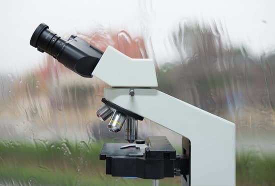
How to find the magnification of a microscope? To figure the total magnification of an image that you are viewing through the microscope is really quite simple. To get the total magnification take the power of the objective (4X, 10X, 40x) and multiply by the power of the eyepiece, usually 10X.
How is the magnification of a microscope calculated? The total magnification of the microscope is calculated from the magnifying power of the objective multiplied by the magnification of the eyepiece and, where applicable, multiplied by intermediate magnifications. … If an object is viewed with the eye from a distance of 250 mm, the magnification is 1x.
How do you find the 100x magnification of a microscope? 100x magnification.
Which microscope is used in metallurgy? The most common metallurgical microscopes are acoustic or ultrasonic microscopes that can be used to examine delimitations, cracks and other anomalies nondestructively and inverted microscopes, which are useful for flat polished metallurgical, ceramic, or optical samples.
How to find the magnification of a microscope? – Related Questions
What is viewed on a scanning tunneling microscope?
A scanning tunneling microscope (STM) is a type of microscope used for imaging surfaces at the atomic level. Its development in 1981 earned its inventors, Gerd Binnig and Heinrich Rohrer, then at IBM Zürich, the Nobel Prize in Physics in 1986. … This means that individual atoms can routinely be imaged and manipulated.
How much does a fluorescence microscope cost?
A fluorescence microscope can cost between $2,400 and $21,000+ depending on the specifications and customizations that you require.
What do blood cells look like under a microscope?
Red blood cells are shaped kind of like donuts that didn’t quite get their hole formed. They’re biconcave discs, a shape that allows them to squeeze through small capillaries. This also provides a high surface area to volume ratio, allowing gases to diffuse effectively in and out of them.
How to focus a dissecting microscope?
Lower the microscope body to its lowest point with the focusing knob on the sides of the microscope arm. Use the focus knob to raise the microscope body until the specimen image is the sharpest.
What are the causes of microscopic hematuria?
The most common causes of microscopic hematuria are urinary tract infection, benign prostatic hyperplasia, and urinary calculi. However, up to 5% of patients with asymptomatic microscopic hematuria are found to have a urinary tract malignancy.
How do you hold a microscope?
Always carry the microscope with 2 hands—place one hand on the microscope arm and the other hand under the microscope base. Do not touch the objective lenses (i.e. the tips of the objectives). Keep the objectives in the scan position and keep the stage low when adding or removing slides.
What regulates light in a microscope?
Iris Diaphragm controls the amount of light reaching the specimen. It is located above the condenser and below the stage. Most high quality microscopes include an Abbe condenser with an iris diaphragm. Combined, they control both the focus and quantity of light applied to the specimen.
Is there a video microscope with hdmi output?
This high-tech while easy to use HDMI Digital Microscope provides 1080p video crystal output directly. It adopts top quality image sensor and corresponding glass microscopic lens, ensuring the best image quality in the digital microscope field.
When carrying the microscope it should be held in a?
WHEN TRANSPORTING THE MICROSCOPE, HOLD IT IN AN UPRIGHT POSITION WITH ONE HAND ON ITS ARM AND THE OTHER SUPPORTING ITS BASE.
How to identify algae under microscope?
Under the microscope they appear like balloon or pear-shaped chrysophycean cells, each with two golden chloroplasts, present in roundish motile colonies. Every cell has two flagella prominent outwards from the colony, and a stem fixed inward near to the colony center.
Did microscopes help discover dna?
Yes, but not in detail. “Many scientists use electron, scanning tunneling and atomic force microscopes to view individual DNA molecules,” said Michael W. Davidson, curator of the National High Magnetic Field Laboratory at Florida State University.
What does the condenser lever do on a microscope?
You will find the condenser under the stage and over the top of the light source. The condenser’s function is to take the light source and narrow the beam to a cone of light to illuminate the specimen to be seen clearly—the condenser and diaphragm of the microscope work in conjunction with each other.
Does microscopic colitis cause a trace of blood in a?
Some patients with microscopic colitis also may report mild abdominal cramps and pain. Blood in the stool is unusual.
What is the smallest lens on a microscope?
Why do you need to start with 4x in magnification on a microscope? The 4x objective lens has the lowest power and, therefore the highest field of view.
What is a microscopic structure in a cell?
Cell organelles are found embedded in the cytoplasm.These are smaller in size and bounded by plasma membrane.
How vinyl records work under a microscope?
As the vinyl disc steadily rotates the stylus moves through the wavy three dimensional grooves. The stylus is a tiny crystal of sapphire or diamond mounted at the very end of a lightweight metal bar like a needle. As the crystal vibrates in the groove, its microscopic bounces are transmitted down the bar.
How can you regulate the diaphragm of a microscope?
Below. The light source is housed in the base of the microscope. It passes through the field iris diaphragm. The size of the field diaphragm is controlled by rotating a knurled ring which is concentric with it.
What are electron microscopes used today?
Electron microscopes are used to investigate the ultrastructure of a wide range of biological and inorganic specimens including microorganisms, cells, large molecules, biopsy samples, metals, and crystals. Industrially, electron microscopes are often used for quality control and failure analysis.
Can you see germs through a microscope?
Bacteria are too small to see without the aid of a microscope. While some eucaryotes, such as protozoa, algae and yeast, can be seen at magnifications of 200X-400X, most bacteria can only be seen with 1000X magnification. … Even with a microscope, bacteria cannot be seen easily unless they are stained.

