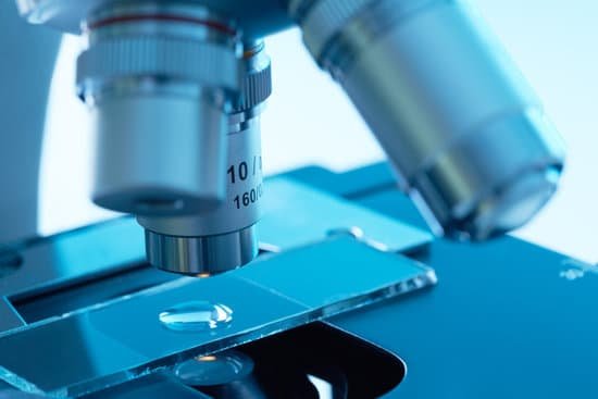
How to remove oil from microscope lens? If you are using a 100x objective with immersion oil, just simply wipe the excess oil off the lens with a kimwipe after use. Occasionally dust may build up on the lightly oiled surface so if you wish to completely remove the oil then you must use an oil soluble solvent.
What is used to clean the oil immersion of microscope? We recommend anhydrous alcohol, a commercially available lens cleaning solution, or blended alcohol. Keep in mind, these cleaning solutions are flammable so you must handle them with care. To help prevent any risks, turn off your microscope and any surrounding lab equipment, and ensure the room is well-ventilated.
What should be used to clean a microscope lens? To clean microscope eyepiece lenses, breathe condensation onto them and then wipe them with lens tissue. Kim-wipes are made by Kleenex and generally will work well. For stubborn spots, wipe the surface with tissue moistened with 95% alcohol. Wipe the lens dry with a dry tissue.
What is the histology of bone tissue? Bone histology.
How to remove oil from microscope lens? – Related Questions
Can microscopic hematuria be normal?
Conclusions. Asymptomatic microscopic hematuria in women is common; however, it is less likely to be associated with urinary tract malignancy among women than men. For women, being older than 60 years, having a history of smoking, and having gross hematuria are the strongest predictors of urologic cancer.
When were microscopes made?
The first compound microscopes date to 1590, but it was the Dutch Antony Van Leeuwenhoek in the mid-seventeenth century who first used them to make discoveries. When the microscope was first invented, it was a novelty item.
Are the functional microscopic units of the kidney?
nephron, functional unit of the kidney, the structure that actually produces urine in the process of removing waste and excess substances from the blood. There are about 1,000,000 nephrons in each human kidney.
How strong of a microscope to see bacteria?
Bacteria are too small to see without the aid of a microscope. While some eucaryotes, such as protozoa, algae and yeast, can be seen at magnifications of 200X-400X, most bacteria can only be seen with 1000X magnification. This requires a 100X oil immersion objective and 10X eyepieces..
What is the piece of glass used with a microscope?
A microscope slide is a thin flat piece of glass, used to hold objects for examination under a microscope. Typically the object is placed or secured (“mounted”) on the slide, and then both are inserted together in the microscope for viewing.
Why are images inverted in a light microscope?
The eyepiece of the microscope contains a 10x magnifying lens, so the 10x objective lens actually magnifies 100 times and the 40x objective lens magnifies 400 times. There are also mirrors in the microscope, which cause images to appear upside down and backwards.
What units are used to measure microscopic objects?
micrometre. micrometre, also called micron, metric unit of measure for length equal to 0.001 mm, or about 0.000039 inch. Its symbol is μm. The micrometre is commonly employed to measure the thickness or diameter of microscopic objects, such as microorganisms and colloidal particles.
What cells can be seen using a light microscope?
You can see yeast cells, animal cells, and plant cells pretty well with a 400x magnification (assuming 10x eyepiece and 40x objective lens). See the image below illustrating the human cheek cells about 80 µm wide (scale bar is 50 µm). There are also many blue speckles outside of the cell.
What type of microscope is used to view algae?
There are two common types of microscopes used in laboratories when studying algae: the compound light microscope (commonly known as a light microscope) and the stereo microscope (commonly known as a dissecting microscope). A light microscope is used to visualize objects flattened onto glass slides in great detail.
How does a pinhole microscope work?
The emitted light passes through the dichroic and is focused onto the pinhole. The light that passes through the pinhole is measured by a detector, ie., a photomultiplier tube. So, there never is a complete image of the sample — at any given instant, only one point of the sample is observed.
What type of microscope uses visible light?
The optical microscope, often referred to as the “light optical microscope,” is a type of microscope that uses visible light and a system of lenses to magnify images of small samples. Optical microscopes are the oldest design of microscope and were possibly designed in their present compound form in the 17th century.
Can a confocal microscope be used for the eyes?
Confocal microscopy allows non-invasive in vivo imaging of all layers of the cornea enabling the clinical investigation of numerous corneal diseases. In this brief review we have evaluated the considerable potential of this powerful technique to undertake detailed morphological analysis of corneal structures.
What can electron microscopes be used for?
Electron microscopy (EM) is a technique for obtaining high resolution images of biological and non-biological specimens. It is used in biomedical research to investigate the detailed structure of tissues, cells, organelles and macromolecular complexes.
What a compound light microscope is used for?
Typically, a compound microscope is used for viewing samples at high magnification (40 – 1000x), which is achieved by the combined effect of two sets of lenses: the ocular lens (in the eyepiece) and the objective lenses (close to the sample).
Can you observe graphene with microscope?
Because graphene on SiC yields uniform samples of relatively large areas, they are well-suited as samples for various types of microscopy. Moreover, because the physical properties of graphene on SiC samples are stable, they may be used as standard reference samples for various material properties.
How many sets of lenses does a compound microscope use?
Typically, a compound microscope is used for viewing samples at high magnification (40 – 1000x), which is achieved by the combined effect of two sets of lenses: the ocular lens (in the eyepiece) and the objective lenses (close to the sample).
Is apple cider vinegar good for microscopic colitis?
For the study, researchers gave the mice apple cider vinegar diluted in drinking water. After one month the scientists found that the vinegar had reduced inflammation in the colon and suppressed proteins that trigger the immune system’s inflammatory response.
What is one disadvantage of using a light microscope?
Disadvantage: Light microscopes have low resolving power. … Electron microscopes are helpful in viewing surface details of a specimen. Disadvantage: Light microscopes can be used only in the presence of light and are costly. Electron microscopes uses short wavelength of electrons and hence have lower magnification.
What is a barlow lens for a microscope?
A Barlow lens is a diverging lens that alters the focal length of a microscope and, therefore, the field of view. They also alter the working distance between the objective lens and the specimen, which is a critical variable for many applications such as PCB soldering and inspection.

