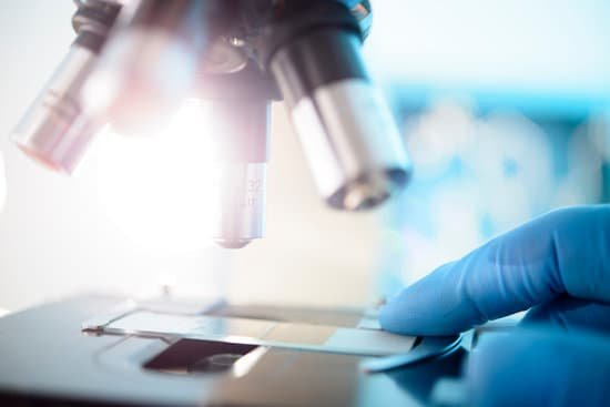
How to use microscope to see blood cells? Place a drop of blood onto a microscope slide. Add a drop of stain to the blood to make the cells easier to see. Carefully place a coverslip over the drop of blood. Sliding it slightly along the microscope slide will spread out the blood cells making them easier to see.
What kind of microscope do you need to see blood cells? Compound microscopes magnify the tiny detail and structure of plant cells, bone marrow and blood cells, single-celled creatures like amoebas, and much more. Almost every homeschool family or hobbyist will need a 400x compound microscope to study cells and tiny organisms in biology and life science.
How do you identify red blood cells under a microscope? They appear as biconcave discs of uniform shape and size (7.2 microns) that lack organelles and granules. Red blood cells have a characteristic pink appearance due to their high content of hemoglobin. The central pale area of each red blood cell is due to the concavity of the disc.
How can you see cells under a microscope? Light microscopy does suffer from a short depth of field at high resolution and this can be seen in the light microscope image of the red blood cells.
How to use microscope to see blood cells? – Related Questions
Do simple microscopes use light?
The optical microscope, also referred to as a light microscope, is a type of microscope that commonly uses visible light and a system of lenses to generate magnified images of small objects. … Basic optical microscopes can be very simple, although many complex designs aim to improve resolution and sample contrast.
What is the principle of confocal microscope?
The basic principle of confocal microscopy is that the illumination and detection optics are focused on the same diffraction-limited spot, which is moved over the sample to build the complete image on the detector.
How to you increase resolving power of a microscope?
One way of increasing the optical resolving power of the microscope is to use immersion liquids between the front lens of the objective and the cover slip. Most objectives in the magnification range between 60x and 100x (and higher) are designed for use with immersion oil.
What do you use to clean microscope lenses?
Dip a lens wipe or cotton swab into distilled water and shake off any excess liquid. Then, wipe the lens using the spiral motion. This should remove all water-soluble dirt.
What did the invention of the microscope significantly advance?
Based on your knowledge of scientific investigations, which stage of scientific investigation did the invention of the microscope significantly advance? Gathering the data for the investigation.
When is oil immersion used in microscope?
Oil immersion objectives are used only at very large magnifications that require high resolving power. Objectives with high power magnification have short focal lengths, facilitating the use of oil. The oil is applied to the specimen (conventional microscope), and the stage is raised, immersing the objective in oil.
Why inverted microscope?
Inverted microscopes are useful for observing living cells or organisms at the bottom of a large container (e.g., a tissue culture flask) under more natural conditions than on a glass slide, as is the case with a conventional microscope.
Why do you need to calibrate a light microscope?
Microscope Calibration can help ensure that the same sample, when assessed with different microscopes, will yield the same results. Even two identical microscopes can have slightly different magnification factors when not calibrated.
Which organelles are visible with the light microscope?
Note: The nucleus, cytoplasm, cell membrane, chloroplasts and cell wall are organelles which can be seen under a light microscope.
What does gram positive look like under a microscope?
Gram positive bacteria have a distinctive purple appearance when observed under a light microscope following Gram staining. This is due to retention of the purple crystal violet stain in the thick peptidoglycan layer of the cell wall.
Can you see bacteria under a light microscope?
Generally speaking, it is theoretically and practically possible to see living and unstained bacteria with compound light microscopes, including those microscopes which are used for educational purposes in schools.
How does the microscope affect the orientation of the image?
The optics of a microscope’s lenses change the orientation of the image that the user sees. A specimen that is right-side up and facing right on the microscope slide will appear upside-down and facing left when viewed through a microscope, and vice versa.
What are the main differences between light and electron microscopes?
Electron microscopes differ from light microscopes in that they produce an image of a specimen by using a beam of electrons rather than a beam of light. Electrons have much a shorter wavelength than visible light, and this allows electron microscopes to produce higher-resolution images than standard light microscopes.
What do the parts of a compound microscope do?
Monocular or Binocular Head: Structural support that holds & connects the eyepieces to the objective lenses. Arm: Supports the microscope head and attaches it to the base. Nosepiece: Holds the objective lenses & attaches them to the microscope head. This part rotates to change which objective lens is active.
What is a comparison microscope and how does it work?
A comparison microscope is a device used to analyze side-by-side specimens. It consists of two microscopes connected by an optical bridge, which results in a split view window enabling two separate objects to be viewed simultaneously.
Are most protist single celled and microscopic?
Protists are a diverse collection of organisms. While exceptions exist, they are primarily microscopic and unicellular, or made up of a single cell. The cells of protists are highly organized with a nucleus and specialized cellular machinery called organelles.
What are the function of base and arm in microscope?
Base – It acts as microscopes support. It also carries microscopic illuminators. Arms – This is the part connecting the base and to the head and the eyepiece tube to the base of the microscope. It gives support to the head of the microscope and it is also used when carrying the microscope.
When would you use a dissecting microscope?
A dissecting microscope is used to view three-dimensional objects and larger specimens, with a maximum magnification of 100x. This type of microscope might be used to study external features on an object or to examine structures not easily mounted onto flat slides.
Can you see different colored cells under microscope?
Using a new technique, researchers have managed to add color information to electron microscopes. Electron microscopes can produce images that are thousands of times more magnified than light microscopes. … Electron microscopes use a beam of electrons to map a sample.

