How would prophase under microscope? During prophase, the molecules of DNA condense, becoming shorter and thicker until they take on the traditional X-shaped appearance. The nuclear envelope breaks down, and the nucleolus disappears. The cytoskeleton also disassembles, and those microtubules form the spindle apparatus.
What does prophase stage look like? In the first phase—prophase—a centriole, located outside the nucleus, divides. The long, threadlike material of the nucleus coils up into visible chromosomes, and the nuclear membrane disappears. From the centrioles, long, thin strands extend in all directions.
What happens in prophase and what does it look like? During prophase, the complex of DNA and proteins contained in the nucleus, known as chromatin, condenses. The chromatin coils and becomes increasingly compact, resulting in the formation of visible chromosomes. Chromosomes are made of a single piece of DNA that is highly organized.
What organelle is visible under a microscope during prophase stage? Chromosomes are not clearly discerned in the nucleus, although a dark spot called the nucleolus may be visible. Prophase. Chromatin in the nucleus begins to condense and becomes visible in the light microscope as chromosomes. The nuclear membrane dissolves, marking the beginning of prometaphase.
How would prophase under microscope? – Related Questions
When was the phase contrast microscope invented?
The easiest and most common way to image biological samples is using phase contrast, which is a special contrast-enhancing imaging method for transmitted-light microscopes invented by Frits Zernike (1888-1966) in 1932 [1] and introduced into microscopic practice by August Köhler (1866-1948) and Loos in 1941 [2, 3].
What organelles cannot be seen under a light microscope?
Some cell parts, including ribosomes, the endoplasmic reticulum, lysosomes, centrioles, and Golgi bodies, cannot be seen with light microscopes because these microscopes cannot achieve a magnification high enough to see these relatively tiny organelles.
How to measure the diameter in a microscope?
Divide the field number by the magnification number to determine the diameter of your microscope’s field of view.
Why must a specimen be thin under the microscope?
A specimen has to be thin so that the light coming from the light source is able to pass through the specimen Specimens are sometimes stained with dyes so that they are easier to distinguish and find.
What are the parts of a microscope slide?
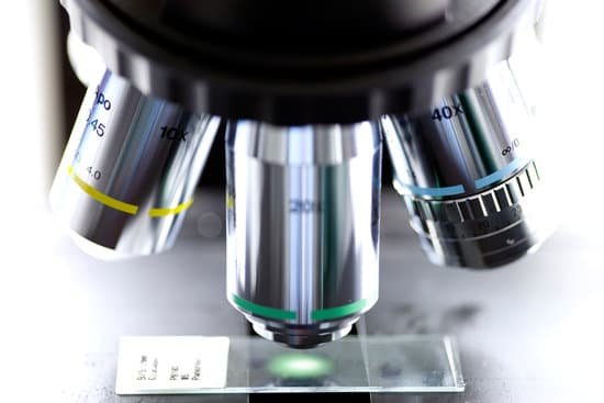
Stage: The flat platform where you place your slides. Stage clips hold the slides in place. Revolving Nosepiece or Turret: This is the part that holds two or more objective lenses and can be rotated to easily change power.
What does microscope mean in history?
A microscope (from Ancient Greek: μικρός mikrós ‘small’ and σκοπεῖν skopeîn ‘to look (at); examine, inspect’) is a laboratory instrument used to examine objects that are too small to be seen by the naked eye.
How do you calculate size via a microscope?
Divide the number of cells in view with the diameter of the field of view to figure the estimated length of the cell. If the number of cells is 50 and the diameter you are observing is 5 millimeters in length, then one cell is 0.1 millimeter long. Measured in microns, the cell would be 1,000 microns in length.
What size microscope to see dust mites?
As I mentioned earlier, dust mites are microscopic creatures which cannot be seen by a naked human eye. However, they can easily be seen under a microscope with at least a 10x magnification lens. Most standard microscopes have 10x magnification eyepieces.
What is the body tube of the microscope?
The microscope body tube separates the objective and the eyepiece and assures continuous alignment of the optics. It is a standardized length, anthropometrically related to the distance between the height of a bench or tabletop (on which the microscope stands) and the position of the seated observer’s…
What microscope achieves the highest magnification and greatest resolution?
The microscope that can achieve the highest magnification and greatest resolution is the electron microscope, which is an optical instrument that is designed to enable us to see microscopic details down to the atomic scale (check also atom microscopy).
Where are omax microscopes made?
The microscope is made in China. The OMAX M82EZ series is the same base microscope, but wi… Q: Does it come put together?
What is a microscope rheostat?
rheostat – alters the current applied to the lamp to control the intensity of the light produced condenser – lens system that aligns and focuses the light from the lamp onto the specimen diaphragms or pinhole apertures – placed in the light path to alter the amount of light that reaches the condenser (for enhancing …
Can flagella be seen with a light microscope?
The basic point about the flagella stain is that the combination of chemicals produces a thickened coat around the flagella, making them more easily seen with a light microscope. Flagella are extremely thin and of small diameter, so they are below the resolution of the light microscope if unstained.
How does one measure objects using a compound light microscope?
Your microscope may be equipped with a scale (called a reticule) that is built into one eyepiece. To measure the length of an object note the number of ocular divisions spanned by the object. … Then multiply by the conversion factor for the magnification used.
What do i use to dry microscope slide for microbiology?
Take a piece of paper towel and hold it close to one edge of the cover slip. This will draw out some water. If too dry, add a drop of water beside the cover slip.
How to correctly focus a microscope?
To focus a microscope, rotate to the lowest-power objective, and place your sample under the stage clips. Play with the magnification using the coarse adjustment knob and move your slide around until it is centered.
How to see onion cells under microscope?
Gently lay a microscopic cover slip on the membrane and press it down gently using a needle to remove air bubbles. Touch a blotting paper on one side of the slide to drain excess iodine/water solution, Place the slide on the microscope stage under low power to observe. Adjust focus for clarity to observe.
How many objective lenses are on the microscope?
Objective Lenses: Usually you will find 3 or 4 objective lenses on a microscope. They almost always consist of 4x, 10x, 40x and 100x powers. When coupled with a 10x (most common) eyepiece lens, total magnification is 40x (4x times 10x), 100x , 400x and 1000x.
Why do scientist need microscopes in laboratory?
Scientists use microscopes to observe objects too small to view with the human eye. Microscopes can magnify an image hundreds of times while…
How many types of microscopes do we have?
There are several different types of microscopes used in light microscopy, and the four most popular types are Compound, Stereo, Digital and the Pocket or handheld microscopes. Some types are best suited for biological applications, where others are best for classroom or personal hobby use.
How to choose a microscope for a child?
Look for a microscope that has glass optics. Many cheap children’s microscopes have plastic eyepieces and objective lenses. Microscopes that do not have glass optics will produce images that are blurry and hard to focus.

