What are the two basic morphological types of microscopic fungi? Fungi can be divided into two basic morphological forms, yeasts and hyphae. Yeastsare unicellular fungi which reproduce asexually by blastoconidia formation (budding) or fission.
What is the basic morphology of fungi? Most fungi are multicellular organisms. They display two distinct morphological stages: the vegetative and reproductive. The vegetative stage consists of a tangle of slender thread-like structures called hyphae (singular, hypha ), whereas the reproductive stage can be more conspicuous. The mass of hyphae is a mycelium.
What are the two different groups of microscopic fungi? Fungal cells are of two basic morphological types: true hyphae (multicellular filamentous fungi) or the yeasts (unicellular fungi), which make pseudohyphae. A fungal cell has a true nucleus, internal cell structures, and a cell wall.
What are the two types of fungal cells? Morphology: Fungi exists in two fundamental forms, filamentous or hyphal form (MOLD) and singe celled or budding form (YEAST). But for the classification of fungi, they are studied as mold, yeast, yeast like fungi and dimorphic fungi.
What are the two basic morphological types of microscopic fungi? – Related Questions
What are the similarities between light and electron microscopes?
Light microscopes and electron microscopes both use radiation – in the form of either light or electron beams, to form larger and more detailed images of objects (e.g. biological specimens, materials, crystal structures, etc.) than the human eye can produce unaided.
How many times can a good light microscope magnify?
Light microscopes allow for magnification of an object approximately up to 400-1000 times depending on whether the high power or oil immersion objective is used. Light microscopes use visible light which passes and bends through the lens system.
What is the definition of electron microscope in science?
Electron microscopy (EM) is a technique for obtaining high resolution images of biological and non-biological specimens. … The transmission electron microscope is used to view thin specimens (tissue sections, molecules, etc) through which electrons can pass generating a projection image.
Are mitochondria visible under a light microscope?
Mitochondria are visible with the light microscope but can’t be seen in detail. Ribosomes are only visible with the electron microscope.
Why does skeletal muscle appear striated under a microscope?
Skeletal muscles are long and cylindrical in appearance; when viewed under a microscope, skeletal muscle tissue has a striped or striated appearance. The striations are caused by the regular arrangement of contractile proteins (actin and myosin).
What is a compound light microscope used for?
Typically, a compound microscope is used for viewing samples at high magnification (40 – 1000x), which is achieved by the combined effect of two sets of lenses: the ocular lens (in the eyepiece) and the objective lenses (close to the sample).
What does a light microscope used to visualise an image?
The light microscope is an instrument for visualizing fine detail of an object. It does this by creating a magnified image through the use of a series of glass lenses, which first focus a beam of light onto or through an object, and convex objective lenses to enlarge the image formed.
What does ebola virus look like under microscope?
The Ebola virus is different: it looks like a strand of spaghetti. And, if you look at an infected cell under an electron microscope, it looks like a ball of spaghetti coming out. Each virus is a long, flexible filament that can adopt different shapes.
Who is the father of compound microscope?
A Dutch father-son team named Hans and Zacharias Janssen invented the first so-called compound microscope in the late 16th century when they discovered that, if they put a lens at the top and bottom of a tube and looked through it, objects on the other end became magnified.
What is the use of a confocal microscope?
The primary functions of a confocal microscope are to produce a point source of light and reject out-of-focus light, which provides the ability to image deep into tissues with high resolution, and optical sectioning for 3D reconstructions of imaged samples.
Why would you use lens paper on a microscope?
Microscope Lens Paper is soft, dust-free paper that is used for cleaning microscope slides and lenses without scratching the glass.
How to use 100x in microscope?
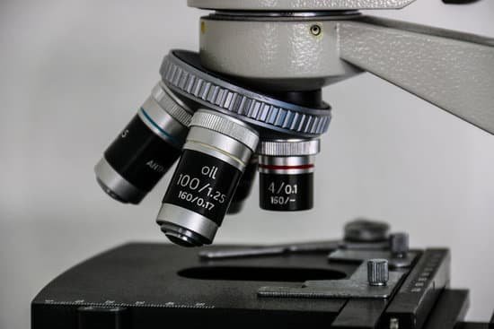
Place a drop of immersion oil on the top of your cover slip and another drop directly on your 100x oil objective lens. Slowly rotate your 100x oil objective lens into place and adjust the fine focus until you get a crisp and clear image.
When was the first microscope invented by robert hooke?
1590: Two Dutch spectacle-makers and father-and-son team, Hans and Zacharias Janssen, create the first microscope. 1667: Robert Hooke’s famous “Micrographia” is published, which outlines Hooke’s various studies using the microscope.
How to calculate power of microscope?
To calculate the total magnification of the compound light microscope multiply the magnification power of the ocular lens by the power of the objective lens. For instance, a 10x ocular and a 40x objective would have a 400x total magnification. The highest total magnification for a compound light microscope is 1000x.
Where did electron microscopes come from?
The invention of the electron microscope by Max Knoll and Ernst Ruska at the Berlin Technische Hochschule in 1931 finally overcame the barrier to higher resolution that had been imposed by the limitations of visible light. Since then resolution has defined the progress of the technology.
Can dna be seen with a microscope?
Given that DNA molecules are found inside the cells, they are too small to be seen with the naked eye. For this reason, a microscope is needed. While it is possible to see the nucleus (containing DNA) using a light microscope, DNA strands/threads can only be viewed using microscopes that allow for higher resolution.
What limits the resolving power of a microscope?
The resolving power of an optical system is ultimately limited by diffraction by the aperture. Thus an optical system cannot form a perfect image of a point.
What is a light microscope used for in science?
light microscopes are used to study living cells and for regular use when relatively low magnification and resolution is enough. electron microscopes provide higher magnifications and higher resolution images but cannot be used to view living cells.
What is microscope slide in biology?
A microscope slide is a thin flat piece of glass, typically 75 by 26 mm (3 by 1 inches) and about 1 mm thick, used to hold objects for examination under a microscope. Typically the object is mounted (secured) on the slide, and then both are inserted together in the microscope for viewing.
How the process of phase contrast works on a microscope?
How phase contrast works. Phase contrast microscopy translates small changes in the phase into changes in amplitude (brightness), which are then seen as differences in image contrast. Unstained specimens that do not absorb light are known as phase objects.
What kind of lens is used in a simple microscope?
A convex lens is used to construct a simple microscope. Convex lens is most widely and popularly used as a reading glass or magnifying glass.

