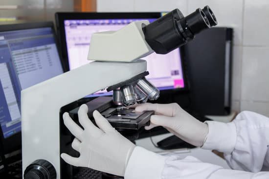
What do we need microscopes for? A microscope is an instrument that is used to magnify small objects. Some microscopes can even be used to observe an object at the cellular level, allowing scientists to see the shape of a cell, its nucleus, mitochondria, and other organelles.
What is the purpose of putting oil on a microscope slide? In light microscopy, oil immersion is a technique used to increase the resolving power of a microscope. This is achieved by immersing both the objective lens and the specimen in a transparent oil of high refractive index, thereby increasing the numerical aperture of the objective lens.
Why is there a need to add oil on the slide prior to viewing the object and 100x magnification? The 100x lens is immersed in a drop of oil placed on the slide in order to eliminate any air gaps and lossof light due to refraction (bending of the light) as the light passes from glass (slide) → air → glass (objective lens). Immersion oil has the same refractive index of glass.
What is the advantage to adding oil to the slide? clear and crisp images. By placing a substance such as immersion oil with a refractive index equal to that of the glass slide in the space filled with air, more light is directed through the objective and a clearer image is observed.
What do we need microscopes for? – Related Questions
Why do chloroplasts move under the microscope?
Chloroplasts migrate in response to different light intensities. Under weak light, chloroplasts gather at an illuminated area to maximize light absorption and photosynthesis rates (the accumulation response). In contrast, chloroplasts escape from strong light to avoid photodamage (the avoidance response).
When was the first microscope invented?
Lens Crafters Circa 1590: Invention of the Microscope. Every major field of science has benefited from the use of some form of microscope, an invention that dates back to the late 16th century and a modest Dutch eyeglass maker named Zacharias Janssen.
How many objective lenses does a microscope have?
Objective Lenses: Usually you will find 3 or 4 objective lenses on a microscope. They almost always consist of 4x, 10x, 40x and 100x powers. When coupled with a 10x (most common) eyepiece lens, total magnification is 40x (4x times 10x), 100x , 400x and 1000x.
What magnifications are possible for the stereo microscope?
The stereo- or dissecting microscope is an optical microscope variant designed for observation with low magnification (2 – 100x) using incident light illumination (light reflected off the surface of the sample is observed by the user), although it can also be combined with transmitted light in some instruments.
Can you see stds under a microscope?
Discharge, tissue, cell or oral fluid sample – Your provider will use a swab to collect samples that will be looked at under a microscope. These samples can test for certain STDs, like chlamydia, gonorrhea, herpes, or HIV.
How to increase contrast of microscope?
Contrast may be improved by placing suitable apertures or filters within the optical path, either in the illuminating system alone (dark ground or Rheinberg illumination), or in conjugate planes in the imaging system (e.g. for phase contrast, differential interference contrast or polarised light microscopy).
What microscope is used to see proteins?
Electron microscope can be used to see protein molecules. An electron microscope that generates high-energy electrons gives an electronic image to observe protein molecules. The high-resolution pictures ensure the arrangement and structure of biomolecules.
When will a compound microscope be used?
Typically, a compound microscope is used for viewing samples at high magnification (40 – 1000x), which is achieved by the combined effect of two sets of lenses: the ocular lens (in the eyepiece) and the objective lenses (close to the sample).
How is total magnification calculated using a light microscope?
To calculate the total magnification of the compound light microscope multiply the magnification power of the ocular lens by the power of the objective lens. For instance, a 10x ocular and a 40x objective would have a 400x total magnification. The highest total magnification for a compound light microscope is 1000x.
What is the difference between macroscopic and microscopic?
The macroscopic level includes anything seen with the naked eye and the microscopic level includes atoms and molecules, things not seen with the naked eye. Both levels describe matter.
What does the prefix micro mean in microscope?
An easy way to remember that the prefix micro- means “small” is through the word microscope, an instrument which allows the viewer to see “small” living things.
What does a condenser lens do on a microscope?
On upright microscopes, the condenser is located beneath the stage and serves to gather wavefronts from the microscope light source and concentrate them into a cone of light that illuminates the specimen with uniform intensity over the entire viewfield.
Where is the ocular on a microscope?
Eyepiece or Ocular is what you look through at the top of the microscope. Typically, standard eyepieces have a magnifying power of 10x. Optional eyepieces of varying powers are available, typically from 5x-30x. Eyepiece Tube holds the eyepieces in place above the objective lens.
Is atomic force microscope expensive?
While it is possible to purchase a simple AFM for as little as a few thousand US dollars, top of the range high-end models can cost half a million dollars or more.
How to carry out malaria test using microscope?
Malaria parasites can be identified by examining under the microscope a drop of the patient’s blood, spread out as a “blood smear” on a microscope slide. Prior to examination, the specimen is stained (most often with the Giemsa stain) to give the parasites a distinctive appearance.
What is the purpose of objectives on a microscope?
Objectives are responsible for primary image formation and play a central role in establishing the quality of images that the microscope is capable of producing. Furthermore, the magnification of a particular specimen and the resolution under which fine specimen detail also heavily depends on microscope objectives.
What is a video microscope?
Video microscopes provide a live feed image directly to a computer, TV or a LCD projector. … The main goal for a video microscope is typically a smooth real-time video image that does not jump and is fluid.
What are binocular microscopes used for?
an instrument used to obtain an enlarged image of small objects and reveal details of structure not otherwise distinguishable. The light path of a darkfield microscope.
When was the invention of the microscope?
In around 1590, Hans and Zacharias Janssen had created a microscope based on lenses in a tube [1]. No observations from these microscopes were published and it was not until Robert Hooke and Antonj van Leeuwenhoek that the microscope, as a scientific instrument, was born.
Is fish oil good for microscopic colitis?
Omega-3 fatty acids, often found in fish oil, are known to have anti-inflammatory properties and several other health benefits. Some people use omega-3 fatty acids to help relieve intestinal inflammation from Crohn’s disease and ulcerative colitis.

