What does anthrax look like under a microscope? Anthrax bacteria have many distinguishing physical characteristics that scientists use to identify them from other types of bacteria under the microscope. For example, the anthrax are shaped like small rods, are non-motile, meaning they do not move around on their own, and are can be stained by certain dyes.
How does anthrax look like? *The characteristic rash of anthrax looks like pink, itchy bumps that occur at the site where B. anthracis comes into contact with scratched or otherwise open skin. The pink bumps progress to blisters, which further progress to open sores with a black base (called an eschar).
What does Bacillus anthracis look like under a microscope? Bacillus anthracis (B. anthracis) is a Gram-positive rod-shaped bacterium that is the causative agent of the disease anthrax. B. anthracis rods typically have dimensions of approximately 1 μm by 4 μm and may occur in chains resembling “boxcars” when observed under a microscope.
How do you identify anthrax? Gram-positive anthrax bacteria (purple rods) in cerebrospinal fluid: If present, a Gram-negative bacterial species would appear pink. (The other cells are white blood cells.)
What does anthrax look like under a microscope? – Related Questions
What plant cell structures are visible under the light microscope?
Note: The nucleus, cytoplasm, cell membrane, chloroplasts and cell wall are organelles which can be seen under a light microscope.
What microscope contains the ocular lens?
Electron microscopes use elec-tron beams instead of light rays, and magnets instead of lenses to observe submicro-scopic particles. This instrument contains two lens systems for magnifying specimens: the ocular lens in the eyepiece and the objective lens located in the nose-piece.
What drugs can cause microscopic colitis?
Your doctor will also ask about any medications you are taking — particularly aspirin, ibuprofen (Advil, Motrin IB, others), naproxen sodium (Aleve), proton pump inhibitors, and selective serotonin reuptake inhibitors (SSRIs) — which may increase your risk of microscopic colitis.
Why were microscopes essential in the discovery of cells?
It allowed scientists to study organisms at the level of their molecules and led to the emergence of the field of cell biology. With the electron microscope, many more cell discoveries were made.
What is the most important part of the microscope?
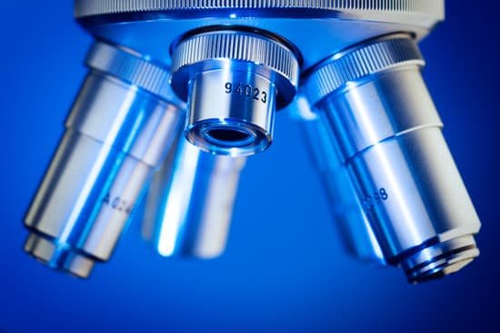
While the modern microscope has many parts, the most important pieces are its lenses. It is through the microscope’s lenses that the image of an object can be magnified and observed in detail.
How do you clean microscope slides?
When slides get soiled, you can clean them with soapy water or isopropyl alcohol. Do not immerse slides in water or soak them in it. This loosens the cover glass adhesive, causing the cover glass to come off and possibly ruin the slide.
How does an stm differ from an optical microscope?
A scanning tunneling microscope (STM) is a non-optical microscope that works by scanning an electrical probe tip over the surface of a sample at a constant spacing. This allows a 3D picture of the surface to be created.
Why does confocal microscope use laser light?
A laser is used to provide the excitation light (in order to get very high intensities). The laser light (blue) reflects off a dichroic mirror. … Our confocal microscope (from Noran) uses a special Acoustic Optical Deflector in place of one of the mirrors, in order to speed up the scanning.
Who created the modern day microscope?
Timeline: under the glass of the microscope. A lot has changed since the late 16th century when father-son scientist duo Hans and Zacharias Janssen invented the world’s first compound microscope.
Is dna microscopic?
Given that DNA molecules are found inside the cells, they are too small to be seen with the naked eye. … While it is possible to see the nucleus (containing DNA) using a light microscope, DNA strands/threads can only be viewed using microscopes that allow for higher resolution.
What is the difference between a sem and tem microscope?
The main difference between SEM and TEM is that SEM creates an image by detecting reflected or knocked-off electrons, while TEM uses transmitted electrons (electrons that are passing through the sample) to create an image.
When was the microscope invented and by whom?
It’s not clear who invented the first microscope, but the Dutch spectacle maker Zacharias Janssen (b. 1585) is credited with making one of the earliest compound microscopes (ones that used two lenses) around 1600. The earliest microscopes could magnify an object up to 20 or 30 times its normal size.
How to enhance contrast on a microscope?
Contrast may be improved by placing suitable apertures or filters within the optical path, either in the illuminating system alone (dark ground or Rheinberg illumination), or in conjugate planes in the imaging system (e.g. for phase contrast, differential interference contrast or polarised light microscopy).
How does the stereo microscope differ from the compound?
One of the main differences between stereo and compound microscopes is the fact that compound microscopes have much higher optical resolution with magnification ranging from about 40x to 1,000x. Stereo microscopes have lower optical resolution power where the magnification typically ranges between 6x and 50x.
Which scientist first observed living cells under a microscope?
Initially discovered by Robert Hooke in 1665, the cell has a rich and interesting history that has ultimately given way to many of today’s scientific advancements.
Can celiac cause microscopic colitis?
Conclusions: Microscopic colitis is more common in patients with celiac disease than in the general population. Patients with celiac disease and microscopic colitis have more severe villous atrophy and frequently require steroids or immunosuppressant therapies to control diarrhea.
How does a measuring microscope work?
Overview. Measuring microscopes combine an optical microscope with a table capable of precise movement to measure targets. As with optical comparators, a telecentric optical system is used to enable accurate measurements. Measurements can be performed in a non-contact manner, so there is no risk of damaging the target.
Do electron microscopes show color of a molecule?
A new method of colorizing electron microscope imagery will make it easier for microbiologists to spot elusive molecules.
How to calculate magnification under a microscope?
To figure the total magnification of an image that you are viewing through the microscope is really quite simple. To get the total magnification take the power of the objective (4X, 10X, 40x) and multiply by the power of the eyepiece, usually 10X.
What’s the purpose of a microscope?
A microscope is an instrument that can be used to observe small objects, even cells. The image of an object is magnified through at least one lens in the microscope. This lens bends light toward the eye and makes an object appear larger than it actually is.
What can you see under an electron microscope?
Electron microscopes are used to investigate the ultrastructure of a wide range of biological and inorganic specimens including microorganisms, cells, large molecules, biopsy samples, metals, and crystals.

