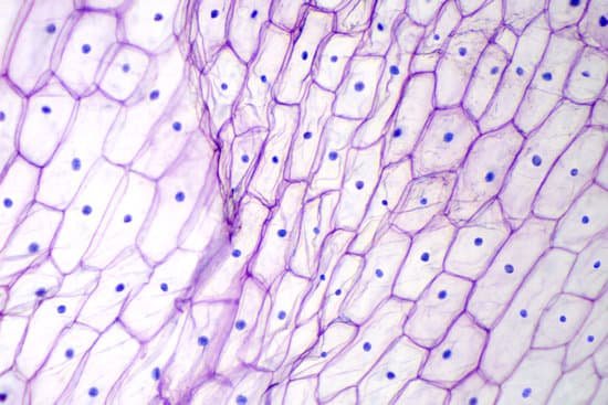
What is one advantage of using a compound light microscope? A compound light microscope is relatively small, therefore it’s easy to use and simple to store, and it comes with its own light source. Moreover, because of their multiple lenses, compound light microscopes are able to reveal a great amount of detail in samples.
What is the advantage of compound light microscope? The advantages of using compound microscope over a simple microscope are: (i) High magnification is achieved, since it uses two lenses instead of one. (ii) It comes with its own light source. (iii) It is relatively small in size; easy to use and simple to handle.
What are 3 advantages of a light microscope? pros and cons
What are the advantages and disadvantages of compound microscope? Resolution: The biggest advantage is that they have a higher resolution and are therefore also able of a higher magnification (up to 2 million times). Light microscopes can show a useful magnification only up to 1000-2000 times. This is a physical limit imposed by the wavelength of the light.
What is one advantage of using a compound light microscope? – Related Questions
What are three uses of electron microscopes?
Electron microscopes are used to investigate the ultrastructure of a wide range of biological and inorganic specimens including microorganisms, cells, large molecules, biopsy samples, metals, and crystals. Industrially, electron microscopes are often used for quality control and failure analysis.
Can you put water on a microscope?
Preparing the slide means to put the pond water onto a microscope slide in a way that it can be viewed through a microscope. First, suck up a small amount of the water in the container with an eye dropper. Then, carefully release the water onto a microscope slide.
What does an atomic force microscope measure?
Atomic-force microscopy (AFM) is a powerful technique that can image almost any type of surface, including polymers, ceramics, composites, glass, and biological samples. AFM is used to measure and localize many forces, including adhesion strength, magnetic forces, and mechanical properties.
Who perfected the microscope?
Zacharias Janssen, credited with inventing the microscope. (Image credit: Public domain.) For millennia, the smallest thing humans could see was about as wide as a human hair. When the microscope was invented around 1590, suddenly we saw a new world of living things in our water, in our food and under our nose.
What type of image does a light microscope produce?
The light microscope is an instrument for visualizing fine detail of an object. It does this by creating a magnified image through the use of a series of glass lenses, which first focus a beam of light onto or through an object, and convex objective lenses to enlarge the image formed.
How do you determine the magnification of a microscope?
It’s very easy to figure out the magnification of your microscope. Simply multiply the magnification of the eyepiece by the magnification of the objective lens. The magnification of both microscope eyepieces and objectives is almost always engraved on the barrel (objective) or top (eyepiece).
What is the lowest magnification of the microscope?
A scanning objective lens provides the lowest magnification power of all objective lenses. 4x is a common magnification for scanning objectives and, when combined with the magnification power of a 10x eyepiece lens, a 4x scanning objective lens gives a total magnification of 40x.
What is the stage adjustment knob on a microscope?
Stage adjustment knobs – located below the stage to control forward/reverse and side to side movement of the stage. Coarse adjustment knob – for focusing ONLY when using scanning objective lens. Fine adjustment knob –brings object into clearest focus.
What do microscopic animals eat?
These minute animals have all the functions of larger creatures: they take in food, excrete wastes, reproduce and communicate. They feed directly on phytoplankton, bacteria and other protozoa. Their respiration releases much of the carbon dioxide incorporated by phytoplankton.
What type of microscope is best for studying dna?
To view the DNA as well as a variety of other protein molecules, an electron microscope is used. Whereas the typical light microscope is only limited to a resolution of about 0.25um, the electron microscope is capable of resolutions of about 0.2 nanometers, which makes it possible to view smaller molecules.
What is the mirror used for on a microscope?
Plane or concave mirror, placed on the microscope base and used to send light onto the specimen and into the microscope optics. The mirror is mounted on a swiveling support, adjusted to reflect natural light or light from an artificial source in the desired direction.
How do you look at bacteria under a microscope?
In order to see bacteria, you will need to view them under the magnification of a microscopes as bacteria are too small to be observed by the naked eye. Most bacteria are 0.2 um in diameter and 2-8 um in length with a number of shapes, ranging from spheres to rods and spirals.
How do you increase resolution on a microscope?
The resolution of a specimen viewed through a microscope can be increased by changing the objective lens. The objective lenses are the lenses that protrude downward over the specimen.
How does a monocular microscope work?
A single tube with interchangeable eyepieces one end, and 1 or more objective lens (often on a revolving turret) the other end. Objects viewed through a monocular microscope will always look flat and without depth. Monocular microscopes are used to study true microscopic sized animals, plants and cells.
How do you use a microscope in agriculture?
Some of the uses of a digital microscope are for visual inspection of seed and grain samples. View your seed and grain samples magnified on a screen instead of an eyepiece to easily perform processes such as varietal identification, seed purity and germination capacity testing.
What does tb look like under a microscope?
Under the microscope, the bacillus is seen as a bright red rod, while the surface that it grows on is colored blue. All bacteria that react in this way to a Ziehl-Neelsen stain are called acid-fast bacteria. The staining technique is used for the diagnosis of TB infection.
Are bacteria visible under microscope?
Bacteria are too small to see without the aid of a microscope. While some eucaryotes, such as protozoa, algae and yeast, can be seen at magnifications of 200X-400X, most bacteria can only be seen with 1000X magnification. … Even with a microscope, bacteria cannot be seen easily unless they are stained.
How to calculate size of specimen microscope?
L, where D is the diameter of the viewing field, E is the estimated number of cells, and L is the length of the cell.
How will the letter e appear placed under a microscope?
The letter “e” appears upside down and backwards under a microscope. Either, diatoms are single celled, or they do not have a cell wall.
Which controls on the microscope affect?
Which controls on the microscope affect the amount of light reaching the ocular lens? The diaphragm and the light intensity adjustment.
What does squamous cell carcinoma look like under a microscope?
At high magnification, this squamous cell carcinoma demonstrates enough differentiation to tell that the cells are of squamous origin. The cells are pink and polygonal in shape with intercellular bridges (seen as desmosomes or “tight junctions” by electron microscopy).

