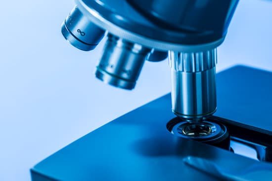
What is the coarse adjustment on a microscope used for? COARSE ADJUSTMENT KNOB — A rapid control which allows for quick focusing by moving the objective lens or stage up and down. It is used for initial focusing.
Why are coarse and fine adjustment used in a microscope? Coarse adjustment knob- Focuses the image under low power (usually the bigger knob) Fine adjustment knob-Sharpens the image under all powers (usually the smaller knob) Arm- supports the body tube and is used to carry the microscope.
When should you use the coarse adjustment on a microscope? There are two knobs on the side of the arm that move the eyepiece. On some microscopes these are located closer to the eyepiece, on others they maybe closer to the stage. The larger knob is called the COARSE ADJUSTMENT and the smaller knob is the FINE ADJUSTMENT. The coarse adjustment is ONLY used on LOW power.
What is the coarse focus on a microscope? Focus (coarse), The coarse focus knob is used to bring the specimen into approximate or near focus. Focus (fine), Use the fine focus knob to sharpen the focus quality of the image after it has been brought into focus with the coarse focus knob.
What is the coarse adjustment on a microscope used for? – Related Questions
What is the greatest resolving power of a light microscope?
The greatest resolving power in optical microscopy is realized with near-ultraviolet light, the shortest effective imaging wavelength. Near-ultraviolet light is followed by blue, then green, and finally red light in the ability to resolve specimen detail.
Is internal kinetic energy microscopic concept?
Internal energy involves energy on the microscopic scale. For an ideal monoatomic gas, this is just the translational kinetic energy of the linear motion of the “hard sphere” type atoms, and the behavior of the system is well described by kinetic theory.
What strength microscope to see sperm?
You can view sperm at 400x magnification. You do NOT want a microscope that advertises anything above 1000x, it is just empty magnification and is unnecessary. In order to examine semen with the microscope you will need depression slides, cover slips, and a biological microscope.
What are microscopic fossils called?
Microfossils are found in rocks and sediments as the microscopic remains of what were once life forms such as plants, animals, fungus, protists, bacteria and archaea. Terrestrial microfossils include pollen and spores. Marine microfossils found in marine sediments are the most common microfossils.
What is the purpose of a fluorescence microscope?
Fluorescence microscopy is highly sensitive, specific, reliable and extensively used by scientists to observe the localization of molecules within cells, and of cells within tissues.
What kind of microscope is a magnifying lens?
Compound Microscope. A simple microscope uses a single lens, so magnifying glasses are simple microscopes. Stereoscopic or dissecting microscopes usually are simple microscopes as well.
How to look through a binocular microscope?
If your microscope is binocular, adjust the interpupillary distance (the distance between the eyepieces) by either sliding or rotating the eyepieces (called adjusting the diopter if present on the microscope) appropriately until you can see only one circle of light with both eyes open.
Who makes laser scanning confocal microscope endoscope?
OptiScan’s laser scanning confocal endomicroscopes are designed for in vivo imaging in medical, pre-clinical and translational applications where conventional confocal microscopes encounter challenges accessing the tissue of interest.
How to identify minerals under microscope?
Several physical properties of minerals can be readily observed from hand specimens, and can be used for recognising and distinguishing different minerals.
What does the iris diaphragm do on a compound microscope?
Iris Diaphragm controls the amount of light reaching the specimen. It is located above the condenser and below the stage. Most high quality microscopes include an Abbe condenser with an iris diaphragm. Combined, they control both the focus and quantity of light applied to the specimen.
Which scientist invented the microscope?
The development of the microscope allowed scientists to make new insights into the body and disease. It’s not clear who invented the first microscope, but the Dutch spectacle maker Zacharias Janssen (b. 1585) is credited with making one of the earliest compound microscopes (ones that used two lenses) around 1600.
How have microscopes improved over time?
Microscopes became more stable and smaller. Lens improvements solved many of the optical problems that were common in earlier versions. The history of the microscope widens and expands from this point with people from around the world working on similar upgrades and lens technology at the same time.
What is medication for microscopic colitis?
The two steroids most often prescribed for microscopic colitis are budesonide (Entocort®) and prednisone. Budesonide is believed to be the safest and most effective medication for treating microscopic colitis.
What microscope lens has the greatest magnification?
The oil immersion objective lens provides the most powerful magnification, with a whopping magnification total of 1000x when combined with a 10x eyepiece.
What is the function of foot in compound microscope?
1. Foot or Base: It is the basal, horse shoe-shaped structure. It provides support to all the remaining parts of the microscope.
Can we see atoms with electron microscope?
“So we can regularly see single atoms and atomic columns.” That’s because electron microscopes use a beam of electrons rather than photons, as you’d find in a regular light microscope. As electrons have a much shorter wavelength than photons, you can get much greater magnification and better resolution.
Can atom be seen by electron microscope?
An electron microscope can be used to magnify things over 500,000 times, enough to see lots of details inside cells. There are several types of electron microscope. A transmission electron microscope can be used to see nanoparticles and atoms.
Where is the diaphragm on the microscope?
The diaphragm can be found near the bottom of the microscope, above the light source and the condenser, and below the specimen stage. This can be controlled through a mechanical lever, or with a dial fitted on the diaphragm.
What industries use scanning electron microscope?
Industries including microelectronics, semiconductors, medical devices, general manufacturing, insurance and litigation support, and food processing, all use scanning electron microscopy as a way to examine the surface composition of components and products.
What is the meaning of electron microscope?
Electron microscopy (EM) is a technique for obtaining high resolution images of biological and non-biological specimens. … The transmission electron microscope is used to view thin specimens (tissue sections, molecules, etc) through which electrons can pass generating a projection image.
What are the two light sources in a dissecting microscope?
Dissecting microscopes utilize two types of light: from incident light (direct illumination) or from transmitted light. Opaque objects placed on the microscope stage can be directly illuminated with incident light from an illuminator.

