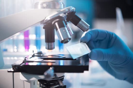
What is the condenser adjustment knob on a microscope? Condenser Focusing Knob – This control is used to precisely adjust the vertical height of the condenser. Condenser Lens – This lens system is located immediately under the stage and focuses the light on the specimen.
What is the function of the condenser on a microscope? On upright microscopes, the condenser is located beneath the stage and serves to gather wavefronts from the microscope light source and concentrate them into a cone of light that illuminates the specimen with uniform intensity over the entire viewfield.
What happens when you adjust the condenser on a microscope? The condenser lens adjustment knob is located below the specimen stage and on the left side. It allows the user to move the condenser lens assembly up or down. As you move the condenser lens up, closer to the specimen, it concentrates (condenses) more light on your specimen.
Where is the condenser height adjustment knob on a microscope? 7. CONDENSER DIAPHRAGM ADJUSTMENT. a. USING THE CONDENSER FOCUS ADJUSTMENT KNOB THAT IS LOCATED ON THE LEFT SIDE OF YOUR MICROSCOPE JUST BELOW THE STAGE, MOVE THE CONDENSER UP TOWARD THE STAGE AS FAR AS IT WILL GO.
What is the condenser adjustment knob on a microscope? – Related Questions
Why do compound light microscopes invert images?
What you would normally classify as a microscope is what you see in a school classroom or on a scientific TV show, and these are called compound microscopes. Compound microscopes invert images! They do this because of the two lenses they have and because of their increased level of magnification.
What is a microscope with only one eyepiece called?
Monocular Microscope. A compound microscope with a single eyepiece. Nosepiece. The upper part of a compound microscope that holds the objective lens. Also called a revolving nosepiece or turret.
What were the first microscopes used for?
1675: Enter Anton van Leeuwenhoek, who used a microscope with one lens to observe insects and other specimen. Leeuwenhoek was the first to observe bacteria.
What is micrometer in microscope?
Microscope micrometers are commonly used for measuring or counting specimens. Eyepiece micrometers (also referred to as “reticles”) are small glass discs with markings on them. The micrometer is mounted in one of the two eyepieces and superimposes an image of the markings over the image of the specimen.
What type of image does a digital microscope produce?
3D measurement is achieved with a digital microscope by image stacking. Using a step motor, the system takes images from the lowest focal plane in the field of view to the highest focal plane. Then it reconstructs these images into a 3D model based on contrast to give a 3D color image of the sample.
How does the first electron microscope work?
The original form of the electron microscope, the transmission electron microscope (TEM), uses a high voltage electron beam to illuminate the specimen and create an image. The electron beam is produced by an electron gun, commonly fitted with a tungsten filament cathode as the electron source.
Can you see the nucleolus under a light microscope?
Thus, light microscopes allow one to visualize cells and their larger components such as nuclei, nucleoli, secretory granules, lysosomes, and large mitochondria. The electron microscope is necessary to see smaller organelles like ribosomes, macromolecular assemblies, and macromolecules.
How did the electron microscope change science?
Implications. The electron microscope ushered in a new era of discoveries printed in academic journals. Atoms were seen by the human eye, as opposed to being merely conceived of. Knowledge of cell structures in plant and animal life increased dramatically as scientists got a first-hand view of the structures themselves …
What are the 4 different types are microscopes?
There are several different types of microscopes used in light microscopy, and the four most popular types are Compound, Stereo, Digital and the Pocket or handheld microscopes. Some types are best suited for biological applications, where others are best for classroom or personal hobby use.
What is an illuminator on a microscope?
Illuminator. There is an illuminator built into the base of most microscopes. The purpose of the illuminator is to provide even, high intensity light at the place of the field aperture, so that light can travel through the condensor to the specimen.
What is the maximum useful magnification for a compound microscope?
The maximum useful magnification for a compound microscope is approximately 1,000 times the N.A. of the objective lens being used. Any effort to increase the total magnification beyond this figure will yield no additional detail.
How tinea capitis is under a microscope?
Microscopy of scalp scrapings or plucked hairs can rapidly confirm the diagnosis of tinea capitis. Specimens are wet-mounted in potassium hydroxide and examined under a light microscope for the presence of hyphae and spores.
How does binocular microscope work?
Many microscopes are binocular and have two ocular lenses. … The oculars have different available magnifications, but usually less than the power of the objective lenses. The objective lenses are at the bottom of the microscope tube nearest the specimen; they gather and focus the light transmitted from the specimen.
Is microscopic hematuria a sign of kidney stones?
The stones are generally painless, so you probably won’t know you have them unless they cause a blockage or are being passed. Then there’s usually no mistaking the symptoms — kidney stones, especially, can cause excruciating pain. Bladder or kidney stones can also cause both gross and microscopic bleeding.
How to calculate total magnification on a light microscope?
To calculate the total magnification of the compound light microscope multiply the magnification power of the ocular lens by the power of the objective lens. For instance, a 10x ocular and a 40x objective would have a 400x total magnification. The highest total magnification for a compound light microscope is 1000x.
What does the condenser lens do on a microscope?
On upright microscopes, the condenser is located beneath the stage and serves to gather wavefronts from the microscope light source and concentrate them into a cone of light that illuminates the specimen with uniform intensity over the entire viewfield.
What is magnifying power of compound microscope?
Compound microscopes typically provide magnification in the range of 40x-1000x, while a stereo microscope will provide magnification of 10x-40x. Compound microscopes are used to view small samples that can not be identified with the naked eye.
What is inside an electron microscope?
The original form of the electron microscope, the transmission electron microscope (TEM), uses a high voltage electron beam to illuminate the specimen and create an image. The electron beam is produced by an electron gun, commonly fitted with a tungsten filament cathode as the electron source.
What is attached to the nosepiece of a microscope?
The revolving nosepiece is the inclined, circular metal plate to which the objective lenses, usually four, are attached. The objective lenses usually provide 4x, 10x, 40x and 100x magnification. The final magnification is the product of the magnification of the ocular and objective lenses.
How to adjust the diopter on a microscope?
Close the eye you just used and look through the other eyepiece with the other eye. Don’t touch the focus knobs, but rather adjust the diopter on the eyepiece if the image is out of focus at all. Move up to the highest magnification objective. Repeat the procedure using the highest objective lens.
Does microscopic colitis cause mucus in stool?
Individuals with irritable bowel syndrome (IBS) do not have colitis, even though this condition is sometimes referred to as having “spastic colitis.” These individuals may have symptoms that mimic colitis such as diarrhea, abdominal pain, and mucus in stool.

