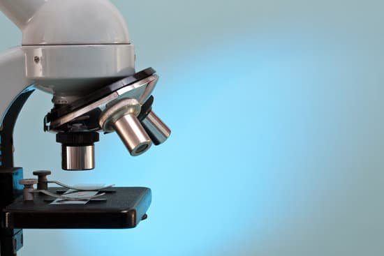
What is the field of view fov in a microscope? Introduction. Microscope field of view (FOV) is the maximum area visible when looking through the microscope eyepiece (eyepiece FOV) or scientific camera (camera FOV), usually quoted as a diameter measurement (Figure 1).
What is the FOV of a 400x microscope? The diameter of field of view (fov) is 0.184 millimeters (184 micrometers). This corresponds to a 0.46 millimeter fov at 400 x magnification.
What is the field size of a microscope? The field number (FN) in microscopy is defined as the diameter of the area in the intermediate image plane that can be observed through the eyepiece. A field number of, e.g., 20 mm indicates that the observed sample area after magnification by the objective lens is restricted to a diameter of 20 mm.
What is the area of a field of view? Field of view (FOV) is the maximum area of a sample that a camera can image. It is related to two things, the focal length of the lens and the sensor size.
What is the field of view fov in a microscope? – Related Questions
What effects do microscopic algae have on corals?
Tiny plant cells called zooxanthellae live within most types of coral polyps. They help the coral survive by providing it with food resulting from photosynthesis. In turn, the coral polyps provide the cells with a protected environment and the nutrients they need to carry out photosynthesis.
How to describe a microscope slide?
A microscope slide is a thin flat piece of glass, typically 75 by 26 mm (3 by 1 inches) and about 1 mm thick, used to hold objects for examination under a microscope. Typically the object is mounted (secured) on the slide, and then both are inserted together in the microscope for viewing.
How to adjust microscope eyepieces?
This is very simple – most microscopes have an adjuster wheel in the centre of the eyepieces to adjust the distance. Otherwise, slide the eyepiece housing to match the width of your eyes. Once you have set this distance, you can then make the diopter adjustment.
Which type of microscope reveals a three dimensional images?
Scanning electron microscopy (SEM) is a powerful technique, traditionally used for imaging the surface of cells, tissues and whole multicellular organisms (see An Introduction to Electron Microscopy for Biologists)(Fig. 1).
What advantages does light microscope have?
The maximum resolution (and therefore magnification) of light microscopes is quite limited compared to electron microscopes – at best, they can magnify up to approximately 2000 times. The advantage of light microscopes (and stereomicroscopes in particular) is that objects can be looked at with little or no preparation.
Why do we use microscopes what are their purpose?
A microscope is an instrument that is used to magnify small objects. Some microscopes can even be used to observe an object at the cellular level, allowing scientists to see the shape of a cell, its nucleus, mitochondria, and other organelles.
What kind of microscope can see atoms?
An electron microscope can be used to magnify things over 500,000 times, enough to see lots of details inside cells. There are several types of electron microscope. A transmission electron microscope can be used to see nanoparticles and atoms.
What is total magnification in light microscope?
To calculate the total magnification of the compound light microscope multiply the magnification power of the ocular lens by the power of the objective lens. For instance, a 10x ocular and a 40x objective would have a 400x total magnification. The highest total magnification for a compound light microscope is 1000x.
What magnification is seen on a stereo microscope?
The stereo- or dissecting microscope is an optical microscope variant designed for observation with low magnification (2 – 100x) using incident light illumination (light reflected off the surface of the sample is observed by the user), although it can also be combined with transmitted light in some instruments.
What is the function of microscope condenser?
On upright microscopes, the condenser is located beneath the stage and serves to gather wavefronts from the microscope light source and concentrate them into a cone of light that illuminates the specimen with uniform intensity over the entire viewfield.
What lenses does a microscope have?
A typical compound microscope will have four objective lenses: one scanning lens, low-power lens, high-power lens, and an oil-immersion lens.
Which procedure extracts tissue for microscopic examination?
Surgical biopsy: The surgical removal of tissue for microscopic examination and diagnosis. Surgical biopsies can be either excisional or incisional.
How to find field of view microscope?
For instance, if your eyepiece reads 10X/22, and the magnification of your objective lens is 40. First, multiply 10 and 40 to get 400. Then divide 22 by 400 to get a FOV diameter of 0.055 millimeters.
What is the structure of a compound microscope?
The three basic, structural components of a compound microscope are the head, base and arm. Arm connects to the base and supports the microscope head. It is also used to carry the microscope.
Can you see dust mites under a microscope?
Dust mites can be difficult to detect due to their small size. These microscopic arthropods are estimated to be only 1/4 to 1/3 millimeters long. You can only see them under a microscope, and even then, they only look like small white spider-like creatures.
What part of the microscope fluorescence?
Typical components of a fluorescence microscope are a light source (xenon arc lamp or mercury-vapor lamp are common; more advanced forms are high-power LEDs and lasers), the excitation filter, the dichroic mirror (or dichroic beamsplitter), and the emission filter (see figure below).
How to clean a dusty microscope?
Put a small amount of lens cleaning fluid or cleaning mixture on the tip of the lens paper. We recommend 70% ethanol because it can effectively and safely clean and disinfect the surface. Larger surfaces, such as a glass plate, may be too large to wipe using this technique.

