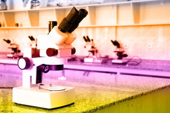
What is the greatest magnification of a light microscope? Using the mathematical equations given above and the values for maximum numerical aperture attainable with the lenses of a light microscope it can be shown that the maximum useful magnification on a light microscope is between 1000X and 1500X. Higher magnification is possible, but resolution will not improve.
How does Clostridium difficile cause colitis? Clostridioides difficile (formerly Clostridium difficile) colitis results from a disturbance of the normal bacterial flora of the colon, colonization by C difficile, and the release of toxins that cause mucosal inflammation and damage. Antibiotic therapy is the key factor that alters the colonic flora.
Can bacteria cause microscopic colitis? It’s unclear what causes microscopic colitis, but researchers believe that it’s likely more than one reason, including infection with bacteria, viruses, parasites, Crohn’s disease, and ulcerative colitis. Your risk of developing microscopic colitis increases with certain genetic predispositions, such as celiac disease.
Can C Diff damage your colon? The C difficile bacterium produces toxins (poisonous substances) that attack the lining of the colon and can cause severe damage to the colon itself. More commonly, C difficile toxins produce diarrhea and abdominal discomfort. Unfortunately, it is resistant to most antibiotics.
What is the greatest magnification of a light microscope? – Related Questions
What is urine microscopic wbc?
If your doctor tests your urine and finds too many leukocytes, it could be a sign of infection. Leukocytes are white blood cells that help your body fight germs. When you have more of these than usual in your urine, it’s often a sign of a problem somewhere in your urinary tract.
Do microscopes bend light?
More advanced microscopes combine multiple lenses to bend light in different ways in order to magnify objects even more. Foldscopes use salt-grain sized lenses that are highly curved. The extreme curvature of these lenses allow for high magnification.
How to use a microscope to look at onion cells?
Gently lay a microscopic cover slip on the membrane and press it down gently using a needle to remove air bubbles. Touch a blotting paper on one side of the slide to drain excess iodine/water solution, Place the slide on the microscope stage under low power to observe. Adjust focus for clarity to observe.
How does an electron microscope detect viruses?
For further characterization immune EM with specific antibodies is used. For diagnostic EM the negative contrast technique is commonly used. The virus particles are surrounded by an electron-dense contrast which reveals the surface structure of the particles.
What is the magnification of zeiss microscope?
The total microscope magnification for visual observation is computed by taking the product of the objective and eyepiece magnifications. For the objective and eyepiece just described, the total lateral magnification would be about 200x (10x eyepiece multiplied by the 20x objective).
What does put under a microscope mean?
: in a state of being watched very closely Celebrities can find it difficult (to be) living under the microscope. The business has recently been put under the microscope by federal investigators.
What is e coli bacteria microscope?
When viewed under the microscope, Gram-negative E. Coli will appear pink in color. The absence of this (of purple color) is indicative of Gram-positive bacteria and the absence of Gram-negative E.
What is the nose piece on a microscope?
1 : the end piece of a microscope body to which an objective is attached and which often consists of a revolving holder for two or more objectives. 2 : the bridge of a pair of eyeglasses.
What does mitochondria look like under a microscope?
Mitochondria are visible under the light microscope although little detail can be seen. Transmission electron microscopy (left) shows the complex internal membrane structure of mitochondria, and electron tomography (right) gives a three-dimensional view.
What is the resolution of the microscope?
In microscopy, the term ‘resolution’ is used to describe the ability of a microscope to distinguish detail. In other words, this is the minimum distance at which two distinct points of a specimen can still be seen – either by the observer or the microscope camera – as separate entities.
What is the function of a eyepiece on a microscope?
The eyepiece, or ocular lens, is the part of the microscope that magnifies the image produced by the microscope’s objective so that it can be seen by the human eye.
What part of a microscope do you look through?
Typically, a compound microscope has one lens in the eyepiece, the part you look through. The eyepiece lens usually magnifies 10 .
What are microscope lenses made of?
Lenses are made of optical glass, a special kind of glass which is much purer and more uniform than ordinary glass. The most important raw material in optical glass is silicon dioxide, which must be more than 99.9% pure.
Which scientists further developed the microscope and when?
In the late 16th century several Dutch lens makers designed devices that magnified objects, but in 1609 Galileo Galilei perfected the first device known as a microscope. Dutch spectacle makers Zaccharias Janssen and Hans Lipperhey are noted as the first men to develop the concept of the compound microscope.
What does increasing gain do in microscope?
The result: higher refresh rates, higher sensitivity and less noise. When viewing objects through the microscope with weak lighting, (for example: when using darkfield or fluorescence applications) the image signal can be enhanced by using the “Gain” slider control in the CapturePro Software.
What shape is leprosy under a microscope?
Leprosy is cause by infection with an intercellular pathogen known as Mycobacterium leprae. M. leprae is a strongly acid-fast, rod-shaped bacterium. It has parallel sides and rounded ends, measuring 1-8 microns in length and 0.2-0.5 micron in diameter, and closely resembles the tubercle bacillus.
What is the resolving power of a transmission electron microscope?
A typical TEM has a resolving power of about 0.2nm. For TEM the typical maximum magnifications is about 1,000,000x. Biological material must be stained with heavy metals to generate contrast in the image. A beam of electrons is scanned over the surface of the specimen.
How to adjust microscope lenses?
Close the eye you just used and look through the other eyepiece with the other eye. Don’t touch the focus knobs, but rather adjust the diopter on the eyepiece if the image is out of focus at all. Move up to the highest magnification objective. Repeat the procedure using the highest objective lens.
Which companies produce transmission electron microscope?
Companies like Bruker Corporation, Danaher Corporation, Asylum Research (Oxford Instruments), Carl Zeiss AG, FEI Co., Danish Micro Engineering, Hitachi High-Technologies Corporation, Jeol Ltd., Nikon Corporation and Olympus Corporation are profiled in this research available at http://www.rnrmarketresearch.com/ …
How does a microscope change an image?
The optics of a microscope’s lenses change the orientation of the image that the user sees. A specimen that is right-side up and facing right on the microscope slide will appear upside-down and facing left when viewed through a microscope, and vice versa.

