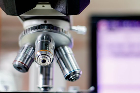
What is the total low power magnification on a microscope? The total magnification of a low power objective lens combined with a 10x eyepiece lens is 100x magnification, giving you a closer view of the slide than a scanning objective lens without getting too close for general viewing purposes.
What is the low power magnification of microscope?
What is the total magnification value for low power? 10X – This objective magnifies the image by a factor of 10 and is referred to as the “low power” objective.
What is the total magnification of a microscope called? 100X (this means that the image being viewed will appear to be 100 times its actual size).
What is the total low power magnification on a microscope? – Related Questions
What do microscopes do?
A microscope is an instrument that can be used to observe small objects, even cells. The image of an object is magnified through at least one lens in the microscope. This lens bends light toward the eye and makes an object appear larger than it actually is.
Can microscopic blood in urine go away?
A blood disease, like sickle cell anemia. A tumor in your urinary tract (may or may not be cancer). Exercise. When this is the cause, hematuria will usually go away in 24 hours.
How to carry a compound microscope?
Always keep your microscope covered when not in use. Always carry a microscope with both hands. Grasp the arm with one hand and place the other hand under the base for support.
Who discovered the scanning electron microscope?
In 1937, Bodo von Borries and Helmut Ruska joined him to develop ways that the principles could be applied, such as to examine biological samples. In the same year, Manfred von Ardenne developed the first scanning electron microscope.
What is the smallest thing measured with a microscope?
Answer 1: The smallest object that we can see using a microscope (in a general sense) is atom, whose size is around 0.1 nano meter. This technique is called Scanning tunneling microscope (STM).
What type of microscope is used in high school classrooms?
Out of the total number of schools, 97 schools are equipped with microscopes, while 6 schools have no microscopes. The most common types of microscopes used in teaching are monocular light microscopes (80%), followed by binocular optical microscopes (16%), digital microscopes (3%), and stereomicroscopes (1%).
What can you see with a microscope?
A microscope is an instrument that is used to magnify small objects. Some microscopes can even be used to observe an object at the cellular level, allowing scientists to see the shape of a cell, its nucleus, mitochondria, and other organelles.
Who discovered by microscope?
Lens Crafters Circa 1590: Invention of the Microscope. Every major field of science has benefited from the use of some form of microscope, an invention that dates back to the late 16th century and a modest Dutch eyeglass maker named Zacharias Janssen.
Where do the specimen goes on a microscope?
All microscopes are designed to include a stage where the specimen (usually mounted onto a glass slide) is placed for observation. Stages are often equipped with a mechanical device that holds the specimen slide in place and can smoothly translate the slide back and forth as well as from side to side.
How much would an electron microscope cost?
The price of a new electron microscope can range from $80,000 to $10,000,000 depending on certain configurations, customizations, components, and resolution, but the average cost of an electron microscope is $294,000. The price of electron microscopes can also vary by type of electron microscope.
What is the purpose of the objectives in a microscope?
Objectives allow microscopes to provide magnified, real images and are, perhaps, the most complex component in a microscope system because of their multi-element design.
Who invented the dissecting microscope?
It was first designed by Cherudin d’Orleans in 1677 by making a small microscope with two separate eyepieces and objective lenses.
Why are electron microscopes better than light microscopes?
Electron microscopes differ from light microscopes in that they produce an image of a specimen by using a beam of electrons rather than a beam of light. Electrons have much a shorter wavelength than visible light, and this allows electron microscopes to produce higher-resolution images than standard light microscopes.
What is the definition of a microscope stage?
Microscope Stages. All microscopes are designed to include a stage where the specimen (usually mounted onto a glass slide) is placed for observation. Stages are often equipped with a mechanical device that holds the specimen slide in place and can smoothly translate the slide back and forth as well as from side to side …
What is microscopic anatomy histology?
Histology, also known as microscopic anatomy or microanatomy, is the branch of biology which studies the microscopic anatomy of biological tissues. Histology is the microscopic counterpart to gross anatomy, which looks at larger structures visible without a microscope.
What is microscopic and macroscopic?
The physical properties of matter can be viewed from either the macroscopic and microscopic level. The macroscopic level includes anything seen with the naked eye and the microscopic level includes atoms and molecules, things not seen with the naked eye. Both levels describe matter.
How are microscopic needles made?
The researchers create the nano-needles in a small ceramic oven. In goes something that looks like aluminium foil with a small burnt patch on it (which is actually a wafer-thin piece of copper), and two hours later at 500 degrees, the copper reacts with oxygen in the heat, creating copper oxide.
Can you see pinworm eggs without a microscope?
If you have pinworms, you might see the worms in the toilet after you go to the bathroom. They look like tiny pieces of white thread. You also might see them on your underwear when you wake up in the morning. But the pinworm eggs are too tiny to be seen without a microscope.
How does immersion oil help with resolution of a microscope?
In light microscopy, oil immersion is a technique used to increase the resolving power of a microscope. This is achieved by immersing both the objective lens and the specimen in a transparent oil of high refractive index, thereby increasing the numerical aperture of the objective lens.
How to connect your digital microscope to your chromebook?
To use your device with a Chromebook, launch the app and then connect the device to the USB port on the Chromebook. If you need to select the digital imager, click on the button just under the gear. Look in the top left corner of the app. You can then select the imager form a drop down list.
What microscope magnification to see bacteria?
While some eucaryotes, such as protozoa, algae and yeast, can be seen at magnifications of 200X-400X, most bacteria can only be seen with 1000X magnification. This requires a 100X oil immersion objective and 10X eyepieces.. Even with a microscope, bacteria cannot be seen easily unless they are stained.

