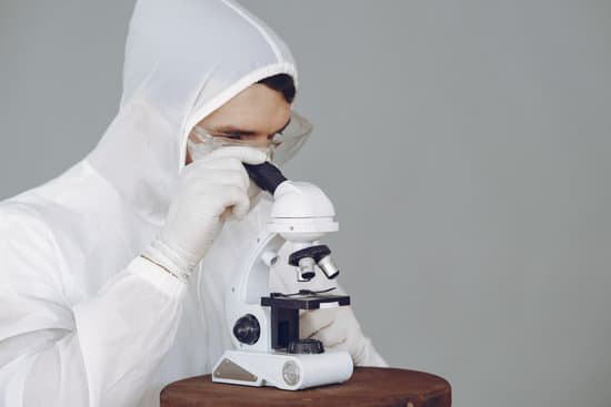
What makes a good microscope camera? In my experience, the parameters that are the most helpful for deciding if a particular camera system will meet your needs are: pixel size, frame rate, quantum efficiency, spectral response, dynamic range, and noise.
How many megapixels do you need for a microscope? Microscope Camera Resolution & Pixels
How do I choose a scientific camera? The key is to find a pixel size that provides sufficient resolution and good signal to noise. Wider fields now allow scientists to capture more cells or image an entire embryo in one view. Speed should depend on the phenomena being captured. Calcium imaging, for example, will require the fastest frame rate possible.
How can you improve the image quality of the microscope? The easiest step to augment image quality is by setting up a cleaning protocol for the microscope and keeping a dust cover over the microscope when it is not in use. A clean and well-maintained microscope will provide significantly better image quality (see Figure 3).
What makes a good microscope camera? – Related Questions
What type of microscope can observe cellular structure?
The light microscope remains a basic tool of cell biologists, with technical improvements allowing the visualization of ever-increasing details of cell structure. Contemporary light microscopes are able to magnify objects up to about a thousand times.
When was the first compound microscope invented?
A Dutch father-son team named Hans and Zacharias Janssen invented the first so-called compound microscope in the late 16th century when they discovered that, if they put a lens at the top and bottom of a tube and looked through it, objects on the other end became magnified.
What does scanning tunneling microscope means?
: a microscope that makes use of the phenomenon of tunneling electrons to map the positions of individual atoms in a surface or to move atoms around on a surface. Other Words from scanning tunneling microscope Example Sentences Learn More About scanning tunneling microscope.
How to deep clean a microscope?
Dip a lens wipe or cotton swab into distilled water and shake off any excess liquid. Then, wipe the lens using the spiral motion. This should remove all water-soluble dirt.
Can you use a light microscope to see bacteria?
Generally speaking, it is theoretically and practically possible to see living and unstained bacteria with compound light microscopes, including those microscopes which are used for educational purposes in schools.
What does skeletal muscle tissue look like under a microscope?
Skeletal muscle looks striped or “striated” – the fibres contain alternating light and dark bands (striations) like horizontal stripes on a rugby shirt. In skeletal muscle, the fibres are packed into regular parallel bundles.
What are microscopes good for when looking at bacteria?
Many affordable microscopes are appropriate for viewing only larger microbes. … On the other hand, compound microscopes are best for looking at all types of microbes down to bacteria. Some, however, are better than others.
What is the function of compound microscope?
Typically, a compound microscope is used for viewing samples at high magnification (40 – 1000x), which is achieved by the combined effect of two sets of lenses: the ocular lens (in the eyepiece) and the objective lenses (close to the sample).
What kind of microscopes have the objectives under the stage?
Inverted Microscope Stages – Inverted microscopes are configured differently than the standard upright microscope. These microscopes have the objectives placed below the stage and use several different condenser configurations to illuminate the specimen.
Why are bacterial cells generally stained for microscopic viewing quizlet?
Why so we stain bacterial cells? … It is used to distinguish between Gram-positive and Gram-negative cells, and allows determination of cell morphology, size, and arrangement. You just studied 7 terms!
What does algae look like under a microscope?
Under the microscope they appear like balloon or pear-shaped chrysophycean cells, each with two golden chloroplasts, present in roundish motile colonies. Every cell has two flagella prominent outwards from the colony, and a stem fixed inward near to the colony center.
What rock is formed by microscopic organisms?
Limestone. Limestone is one of the most widespread sedimentary rocks. Many organisms, from corals to microscopic foraminifera, grow shells composed of carbonates. Most limestone forms when these organisms die and their carbonate shells accumulate in shallow seas.
What does a parfocal microscope do?
Parfocal means that when one objective lens is in focus, then the other objectives will also be in focus. … Parfocal means that the microscope is self-cleaning and needs no maintenance.
Can we see molecules structure under microscope?
This, believe it or not, is a microscope. It can help us see very small particles like molecules by feeling the particle with the tip of its needle. These very powerful microscopes are called atomic force microscopes, because they can see things by feeling the forces between atoms. …
How do you calculate the power of your microscope?
It’s very easy to figure out the magnification of your microscope. Simply multiply the magnification of the eyepiece by the magnification of the objective lens. The magnification of both microscope eyepieces and objectives is almost always engraved on the barrel (objective) or top (eyepiece).
Why do light microscopes have poor resolution?
The resolution of an image is limited by the wavelength of radiation used to view the sample. … The wavelength of light is much larger than the wavelength of electrons, so the resolution of the light microscope is a lot lower.
What is the resolving power of electron microscope?
The resolution limit of electron microscopes is about 0.2nm, the maximum useful magnification an electron microscope can provide is about 1,000,000x.
Is archaea microscopic?
Archaea are a group of microscopic organisms that were discovered in the early 1970s. Like bacteria, they are single-celled prokaryotes. Archaeans were originally thought to be bacteria until DNA analysis showed that they are different organisms.
What is the history of microscope?
In the late 16th century several Dutch lens makers designed devices that magnified objects, but in 1609 Galileo Galilei perfected the first device known as a microscope. Dutch spectacle makers Zaccharias Janssen and Hans Lipperhey are noted as the first men to develop the concept of the compound microscope.
How to measure the size of something under a microscope?
Divide the number of cells in view with the diameter of the field of view to figure the estimated length of the cell. If the number of cells is 50 and the diameter you are observing is 5 millimeters in length, then one cell is 0.1 millimeter long. Measured in microns, the cell would be 1,000 microns in length.
What are microscopes used for in biology?
A microscope is an instrument that is used to magnify small objects. Some microscopes can even be used to observe an object at the cellular level, allowing scientists to see the shape of a cell, its nucleus, mitochondria, and other organelles.

