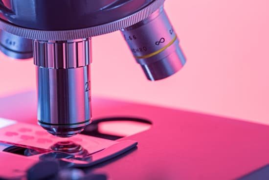
What microscope can see dna molecule? To view the DNA as well as a variety of other protein molecules, an electron microscope is used. Whereas the typical light microscope is only limited to a resolution of about 0.25um, the electron microscope is capable of resolutions of about 0.2 nanometers, which makes it possible to view smaller molecules.
Can TEM microscopes see DNA? Although DNA is visible when observed with the electron microscope, the resolution of the image obtained is not high enough to allow for deciphering the sequence of the individual bases, i.e., DNA sequencing.
What magnification do you need to see DNA? A stain has been used to dye DNA in the cells. This will make the chromosomes show up in a darker color than the rest of the cell. Focusing the microscope with 40x objective should give you a close enough view of the chromosomes to find each phase.
What is DNA microscopy? DNA microscopy is an optics-free imaging method based on chemical reactions and a computational algorithm to infer spatial organization of transcripts while simultaneously preserving full sequence information. DNA MICROSCOPY.
What microscope can see dna molecule? – Related Questions
What does compound microscope mean in science?
A compound microscope is a microscope that uses multiple lenses to enlarge the image of a sample. … The total magnification is calculated by multiplying the magnification of the ocular lens by the magnification of the objective lens. Light is passed through the sample (called transmitted light illumination).
How much does a compound microscope cost?
The most popular compound microscopes from some of the most well-known brands cost on average around $900-$1,200, although there are beginner microscopes that are just above the toy level that cost $100.
What can only be seen with an electron microscope?
Mitochondria are visible with the light microscope but can’t be seen in detail. Ribosomes are only visible with the electron microscope.
What does telophase look like under a microscope?
During the last of the mitosis phases, telophase, the spindle fibers disappear and the cell membrane forms between the two sides of the cell. … The new nucleoli may be visible, and you will note a cell membrane (or cell wall) between the two daughter cells.
Why is the electron microscope better than the regular one?
Electrons have much a shorter wavelength than visible light, and this allows electron microscopes to produce higher-resolution images than standard light microscopes. Electron microscopes can be used to examine not just whole cells, but also the subcellular structures and compartments within them.
Why are things more detail under a microscope?
Microscopes enhance our sense of sight – they allow us to look directly at things that are far too small to view with the naked eye. Microscopes increase the amount of detail we can see. …
What does a scanning electron microscope look at?
A scanning electron microscope (SEM) scans a focused electron beam over a surface to create an image. The electrons in the beam interact with the sample, producing various signals that can be used to obtain information about the surface topography and composition.
What can be seen in light microscope?
Thus, light microscopes allow one to visualize cells and their larger components such as nuclei, nucleoli, secretory granules, lysosomes, and large mitochondria. The electron microscope is necessary to see smaller organelles like ribosomes, macromolecular assemblies, and macromolecules.
How does the compound microscope magnify an object?
When light reflects off of an object being viewed under the microscope and passes through the lens, it bends towards the eye. This makes the object look bigger than it actually is.
What is the most commonly used microscope in a clinic?
One of the most common microscopes is the light microscope. These use light to illuminate an image, while one or sometimes multiple lenses magnify the specimen.
How do you find the maximum magnification of a microscope?
Light microscopes combine the magnification of the eyepiece and an objective lens. Calculate the magnification by multiplying the eyepiece magnification (usually 10x) by the objective magnification (usually 4x, 10x or 40x). The maximum useful magnification of a light microscope is 1,500x.
What is compound microscope in physics?
A compound microscope consists of two lenses, an objective lens (close to the object) and an eye lens (close to the eye). … The objective lens forms a real image of the object close to the eye lens, from which the eye lens gives a greatly magnified virtual image.
Which microscope objective is best for studying bacteria?
Which of the microscope objectives is most satisfactory for studying bacteria in stained preparations? In wet-mount preparations? Why? Usually 100X (oil immersion) lens is used for stained preps.
What holds the slide in place while on the microscope?
Stage clips hold the slides in place. Revolving Nosepiece or Turret: This is the part that holds two or more objective lenses and can be rotated to easily change power. Objective Lenses: Usually you will find 3 or 4 objective lenses on a microscope.
What does bacillus subtilis look like under a microscope?
Bacillus subtilis is rod-shaped and typically 4-10 microns long. … Bacillus Subtilis captured under the U2 biological microscope at 40x. Bacillus subtilis is considered one of the best studied gram-positive bacterium and a model organism for studying bacterial chromosome replication and cell differentiation.
How to put fungi under microscope?
All you have to do is to place the fungus, fertile side downwards, on to a microscope slide and wait an hour or two. Unlike when trying to make a nice spore print you don’t even need to cover everything over, although I generally do just as a reminder that there is a sharp-edged piece of glass underneath.
How light travels through a compound microscope?
Light from a mirror is reflected up through the specimen, or object to be viewed, into the powerful objective lens, which produces the first magnification. The image produced by the objective lens is then magnified again by the eyepiece lens, which acts as a simple magnifying glass.
Why is a microscope so important to forensic?
When it comes to solving a crime, even trace evidence may make or break a case. For this reason, microscopes are essential for many investigative purposes, because they can magnify an object to such great detail. … Microscopes can also be used to compare hairs, fibers or other particulates recovered from the scene.
When was the transmission electron microscope first used?
The first TEM was demonstrated by Max Knoll and Ernst Ruska in 1931, with this group developing the first TEM with resolution greater than that of light in 1933 and the first commercial TEM in 1939. In 1986, Ruska was awarded the Nobel Prize in physics for the development of transmission electron microscopy.
What was looked at with the first microscope?
He saw bacteria, yeast, blood cells and many tiny animals swimming about in a drop of water. From his great contributions, many discoveries and research papers, Anthony Leeuwenhoek (1632-1723) has since been called the “Father of Microscopy”.
How many ocular lenses does a monocular compound microscope have?
Monocular – only use one eyepiece when viewing the specimen. You are restricted if you want to use a CCD camera because this would occupy the eyepiece. However, monocular microscopes are light weight and are inexpensive.

