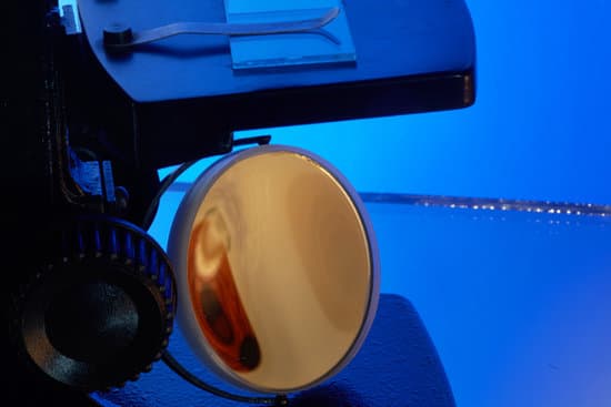
What microscopes are used in schools? The most common types of microscopes used in teaching are monocular light microscopes (80%), followed by binocular optical microscopes (16%), digital microscopes (3%), and stereomicroscopes (1%). A total of 43% of teachers perform microscopy using the demonstration method, and 37% of teachers use practical work.
What microscopes do most schools use? Five Best Microscopes for College & High School Students
What type of microscope is used in schools and why? Compound microscopes are usually used with transmitted light to look through transparent specimens; the useful school magnification range is 10-400x. The image is inverted.
What is the main function of the draw tube on a microscope? The drawtube (if present) carries the ocular, it can be adjusted to control tube length and so effect corrections for the objective lens. The drawtube may also be convenient in calibrating an eyepiece-micrometre by allowing a minor magnification change to simplify the conversion factor.
What microscopes are used in schools? – Related Questions
Are there microscopic bugs on your eyelashes?
Eyelash mites are tiny cigar-shaped bugs found in bunches at the base of your eyelashes. They’re normal and usually harmless, unless you have too many of them. Also known as demodex, each mite has four pairs of legs that make it easy to grip tube-shaped things — like your lashes.
What are the types of microscope and their uses?
There are several different types of microscopes used in light microscopy, and the four most popular types are Compound, Stereo, Digital and the Pocket or handheld microscopes. Some types are best suited for biological applications, where others are best for classroom or personal hobby use.
What causes microscopic polyangiitis?
Certain genes may be a cause. Problems with the immune system may also be a cause. MPA sometimes happens along with an autoimmune disease, such as rheumatoid arthritis (RA). An autoimmune disease is caused by a problem with the immune system.
What type of microscope is used to observe living cells?
The light microscope remains a basic tool of cell biologists, with technical improvements allowing the visualization of ever-increasing details of cell structure. Contemporary light microscopes are able to magnify objects up to about a thousand times.
What is the stage in a microscope?
All microscopes are designed to include a stage where the specimen (usually mounted onto a glass slide) is placed for observation. Stages are often equipped with a mechanical device that holds the specimen slide in place and can smoothly translate the slide back and forth as well as from side to side.
When did people discover microscopic diseases?
The existence of microscopic organisms was discovered during the period 1665-83 by two Fellows of The Royal Society, Robert Hooke and Antoni van Leeuwenhoek.
What magnification does an electron microscope have?
An electron microscope, on the other hand, uses a beam of electrons rather than light to form the image. The magnification of an electron microscope may be as high as 10,000,000x, with a resolution of 50 picometers (0.05 nanometers).
Why is the image in the microscope inverted?
As we mentioned above, an image is inverted because it goes through two lens systems, and because of the reflection of light rays. The two lenses it goes through are the ocular lens and the objective lens. An ocular lens is the one closest to your eye when looking through a microscope or telescope.
Why is it desirable that microscope objectives be parfocal?
Why is it desirable that microscope objective be parfocal? So that you do not have to refocus the microscope every time you switch lenses. … Which controls on the microscope affect the amount of light reaching the ocular lens? The diaphragm and the light intensity adjustment.
Does looking at a microscope make you nauseous?
You may feel sick from the motion of cars, airplanes, trains, amusement park rides, or boats or ships. You could also get sick from video games, flight simulators, or looking through a microscope. In these cases, your eyes see motion, but your body doesn’t sense it.
Who invented the first crude microscope by grinding glass?
Grinding glass to use for spectacles and magnifying glasses was commonplace during the 13th century. In the late 16th century several Dutch lens makers designed devices that magnified objects, but in 1609 Galileo Galilei perfected the first device known as a microscope.
Can a compound microscope view the mitochondria of a cell?
Mitochondria are visible with the light microscope but can’t be seen in detail. Ribosomes are only visible with the electron microscope.
What does quartz look like under a microscope?
Under the microscope, quartz lacks cleavage and colour and has low first-order, grey-white interference colours (Figure 53b and c). Being chemically stable, quartz crystals look clean compared with feldspars, which are almost always turbid or cloudy.
Does interstitial cystitis cause microscopic hematuria?
Interstitial cystitis (IC) is an ill-defined, chronic inflammatory condition of the bladder of unknown etiology characterized by pelvic pain, frequency, urgency, and nocturia. These patients classically present with a constellation of urologic complaints, which may include microscopic and gross hematuria.
What are the parts of microscope and each function?
Eyepiece Lens: the lens at the top that you look through, usually 10x or 15x power. Tube: Connects the eyepiece to the objective lenses. Arm: Supports the tube and connects it to the base. Base: The bottom of the microscope, used for support.
What microscope for apologia biology?
This National Optical-brand, model 131 – CLED microscope is designed to meet or exceed high school teaching requirements, is ruggedly built and includes all the teaching essentials plus popular “student proofing” features. This microscope can be used with biology and advanced biology.
How microscopes changed the world?
The invention of the microscope allowed scientists and scholars to study the microscopic creatures in the world around them. … The microscope allowed human beings to step out of the world controlled by things unseen and into a world where the agents that caused disease were visible, named and, over time, prevented.
Who is most associated with the early use of microscopes?
The first compound microscopes date to 1590, but it was the Dutch Antony Van Leeuwenhoek in the mid-seventeenth century who first used them to make discoveries.
How do you calculate magnification on a light microscope?
To calculate the total magnification of the compound light microscope multiply the magnification power of the ocular lens by the power of the objective lens. For instance, a 10x ocular and a 40x objective would have a 400x total magnification. The highest total magnification for a compound light microscope is 1000x.

