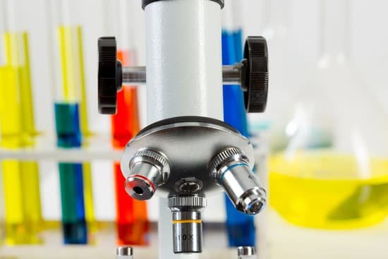
What would you use an electron microscope for? Electron microscopy (EM) is a technique for obtaining high resolution images of biological and non-biological specimens. It is used in biomedical research to investigate the detailed structure of tissues, cells, organelles and macromolecular complexes.
What is an electron microscope best used for? Electron microscopes are used to investigate the ultrastructure of a wide range of biological and inorganic specimens including microorganisms, cells, large molecules, biopsy samples, metals, and crystals. Industrially, electron microscopes are often used for quality control and failure analysis.
What can be seen with electron microscope? Some electron microscopes can detect objects that are approximately one-twentieth of a nanometre (10-9 m) in size – they can be used to visualise objects as small as viruses, molecules or even individual atoms.
What does the electron microscope used to work? The electron microscope uses a beam of electrons and their wave-like characteristics to magnify an object’s image, unlike the optical microscope that uses visible light to magnify images. … This beam is focused onto the sample using a magnetic lens.
What would you use an electron microscope for? – Related Questions
What are the two knobs on a microscope called?
There are two knobs on the side of the arm that move the eyepiece. On some microscopes these are located closer to the eyepiece, on others they maybe closer to the stage. The larger knob is called the COARSE ADJUSTMENT and the smaller knob is the FINE ADJUSTMENT. The coarse adjustment is ONLY used on LOW power.
Is microscopic hematuria a sign of cancer?
This is called “microscopic hematuria,” and it can only be found with a urine test. General urine tests are not used to make a specific diagnosis of bladder cancer because hematuria can be a sign of several other conditions that are not cancer, such as an infection or kidney stones.
What is microscopic hematuria mean?
Microscopic hematuria means that the blood can only be seen with a microscope. Gross hematuria means the urine appears red or the color of tea or cola to the naked eye.
How do you change the magnification of a microscope?
On a standard stereo microscope (not a common main objective stereo microscope) the objective lens is built into the microscope and the only way to change this magnification is by adding an auxiliary lens to the existing objective lens. These are typically available in increments of 0.5x, 0.75x and 1.5x magnification.
Why use microscopes?
A microscope lets the user see the tiniest parts of our world: microbes, small structures within larger objects and even the molecules that are the building blocks of all matter. The ability to see otherwise invisible things enriches our lives on many levels.
What does optical microscope do?
The optical microscope, often referred to as the “light optical microscope,” is a type of microscope that uses visible light and a system of lenses to magnify images of small samples. Optical microscopes are the oldest design of microscope and were possibly designed in their present compound form in the 17th century.
How to clean microscope lens objective?
Place the objective lens on a dust-free surface. 2. Gently blow away loose dust that is on the surface of the optical glass with a dust blower, as if any dust left on throughout the cleaning process could scratch the optical glass or coating. Blow the air across the lens surface to avoid damaging it.
Do gynecologists use microscopes?
Cardiothoracic and pediatric surgeons tend only to utilize loupes, whereas neurosurgeons tend only to use microscopes. General surgeons, urologists, orthopedic surgeons, and gynecologists are infrequent users or nonusers of magnification, and when required will utilize loupes rather than microscopes.
How does a petrographic microscope work?
In the petrographic microscope, the light is collimated by the condenser into a bundle of beams,all parallel to the optical aids of the microscope. … The light beams are polarized in one direction (by the polarizer) before the light reaches the specimen. This light is called plane polarized light.
What is a base on a microscope?
Base: The bottom of the microscope, used for support. … If your microscope has a mirror, it is used to reflect light from an external light source up through the bottom of the stage.
What is a metallographic microscope?
Metallographic microscopes are used to identify defects in metal surfaces, to determine the crystal grain boundaries in metal alloys, and to study rocks and minerals. This type of microscope employs vertical illumination, in which the light source is inserted into the microscope tube…
Are kim wipes for a microscope?
Kimwipes continues to provide the high-performance wiper solutions you need to clean microscopes, equipment and lab surfaces, parts, instruments and lenses. They also can be used to wipe small quantities of solvents or other liquids from hands, tools, equipment or other surfaces.
What is the most powerful microscope in the world?
Lawrence Berkeley National Labs just turned on a $27 million electron microscope. Its ability to make images to a resolution of half the width of a hydrogen atom makes it the most powerful microscope in the world.
What are the slides called for a microscope?
A glass slide is a thin, flat, rectangular piece of glass that is used as a platform for microscopic specimen observation. A typical glass slide usually measures 25 mm wide by 75 mm, or 1 inch by 3 inches long, and is designed to fit under the stage clips on a microscope stage.
How much can a compound light microscope magnify objects?
Compound microscopes typically provide magnification in the range of 40x-1000x, while a stereo microscope will provide magnification of 10x-40x. Compound microscopes are used to view small samples that can not be identified with the naked eye.
How does saccharomyces microscopically differ from other specimens?
Saccharomyces is a yeast, a single-celled fungus. How does Saccharomyces microscopically differ from the other specimens? The bacteria were all small, pink clusters of cells with various shapes and sizes. The Saccharomyces was similar in appearance but were blue and slightly larger than the bacteria.
What is the shortest objective on a microscope called?
After the light has passed through the specimen, it enters the objective lens (often called “objective” for short). The shortest of the three objectives is the scanning-power objective lens (N), and has a power of 4X.
Why can t you see ribosomes under a light microscope?
Some cell parts, including ribosomes, the endoplasmic reticulum, lysosomes, centrioles, and Golgi bodies, cannot be seen with light microscopes because these microscopes cannot achieve a magnification high enough to see these relatively tiny organelles.
What is the disadvantage of a light microscope?
Disadvantage: Light microscopes have low resolving power. … Electron microscopes are helpful in viewing surface details of a specimen. Disadvantage: Light microscopes can be used only in the presence of light and are costly. Electron microscopes uses short wavelength of electrons and hence have lower magnification.
How to estimate size of specimen under microscope?
Divide the number of cells in view with the diameter of the field of view to figure the estimated length of the cell. If the number of cells is 50 and the diameter you are observing is 5 millimeters in length, then one cell is 0.1 millimeter long. Measured in microns, the cell would be 1,000 microns in length.
What do you see under a light microscope during anaphase?
If you view early anaphase using a microscope, you will see the chromosomes clearly separating into two groups. If you are looking at late anaphase, these groups of chromosomes will be on opposite sides of the cell.

