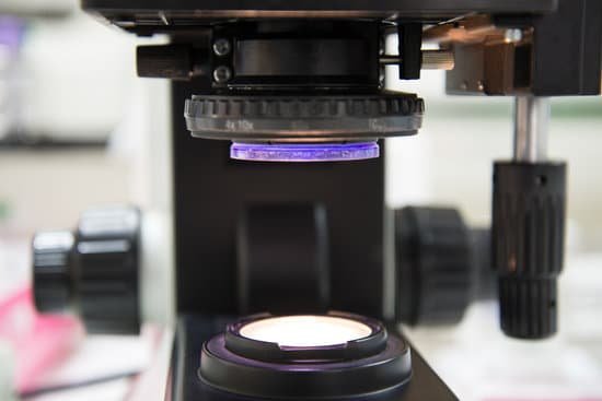
What would you view in a microscope? A microscope is an instrument that is used to magnify small objects. Some microscopes can even be used to observe an object at the cellular level, allowing scientists to see the shape of a cell, its nucleus, mitochondria, and other organelles.
What does a scanning electron microscope show? Because of its great depth of focus, a scanning electron microscope is the EM analog of a stereo light microscope. It provides detailed images of the surfaces of cells and whole organisms that are not possible by TEM. It can also be used for particle counting and size determination, and for process control.
What do SEM images show? A scanning electron microscope (SEM) is a type of microscope which uses a focused beam of electrons to scan a surface of a sample to create a high resolution image. SEM produces images that can show information on a material’s surface composition and topography.
What is SEM analysis used for? Scanning Electron Microscopy, or SEM analysis, provides high-resolution imaging useful for evaluating various materials for surface fractures, flaws, contaminants or corrosion.
What would you view in a microscope? – Related Questions
Why do biologists use microscopes?
Microscopes are the backbone of studying biology. The biologists use them to view the details that cannot be seen by the naked eye such as the small parasites and small organisms which is important for the disease control research.
What kind of bug is almost microscopic and yellow?
The yellow ones are the tiniest, and they’re known as Frankliniella Occidentalis. These tiny thrips are plant bugs and grow up to 1/50th of an inch long.
Who found first microscope?
Lens Crafters Circa 1590: Invention of the Microscope. Every major field of science has benefited from the use of some form of microscope, an invention that dates back to the late 16th century and a modest Dutch eyeglass maker named Zacharias Janssen.
What is the use of eyepiece lens in microscope?
The eyepiece, or ocular lens, is the part of the microscope that magnifies the image produced by the microscope’s objective so that it can be seen by the human eye.
What the letter e looks like under a microscope?
The letter “e” appears upside down and backwards under a microscope. Either, diatoms are single celled, or they do not have a cell wall.
How to disinfect microscope slides?
This can be dish washing fluid, or it can be a more specialized cleaning solution for slides, such as an ethyl alcohol solution. Apply the soap uniformly across both sides of the glass with something that won’t scratch the slide, such as a lint-free microfiber towel. Rinse the slide thoroughly using warm running water.
Who invented the microscope and how?
In the late 16th century several Dutch lens makers designed devices that magnified objects, but in 1609 Galileo Galilei perfected the first device known as a microscope. Dutch spectacle makers Zaccharias Janssen and Hans Lipperhey are noted as the first men to develop the concept of the compound microscope.
Why is a compound microscope called a compound?
The term “compound” in compound microscopes refers to the microscope having more than one lens. Devised with a system of combination of lenses, a compound microscope consists of two optical parts, namely the objective lens and the ocular lens.
What does stomata look like under a microscope?
When viewed under the microscope, it’s possible to see the epidermal cells that tend to be irregular. In addition to the epidermal cells, one will also see the leaf spores (stomata) in between the epidermal cells. Typically, the stomata are bean shaped and will appear denser (darker) under the microscope.
Which science invented the microscope?
In the late 16th century several Dutch lens makers designed devices that magnified objects, but in 1609 Galileo Galilei perfected the first device known as a microscope. Dutch spectacle makers Zaccharias Janssen and Hans Lipperhey are noted as the first men to develop the concept of the compound microscope.
How to turn on a microscope?
Place your microscope on a flat surface and connect its power cord into an outlet. Now, flip on the light switch, which is typically located on the bottom of the microscope. After flipping the switch, the light should come out of the illuminator, which is the light source.
What can you see with a simple microscope?
It is usually used for the study of microscopic algae, fungi, and biological specimens. It is commonly used by watchmakers to see the magnified view of small parts of a watch. It is also used by the jewelers to see the magnified view of the fine parts of jewelry.
Do scientists use a microscope to see archaea?
The small size of Archaea is an obstacle to their detailed structural study and generally precludes light microscopy. … Archaea must be prepared in some manner before imaging to withstand the rigors of the vacuum of the electron microscope.
Do sperm cells move under a microscope?
New research upends more than three centuries of beliefs about how sperm move. Under a microscope, human sperm seem to swim like wiggling eels, tails gyrating to and fro as they seek an egg to fertilize.
What is the field of view on a compound microscope?
Field of view microscope definition in simple terms it is the area you see under the microscope for a particular magnification. Say, for example, you are viewing a cell or specimen under an optical microscope. The diameter of the circle that you see is the field of view of the microscope.
What happens to an image under a microscope?
Microscopes invert images which makes the picture appear to be upside down. The reason this happens is that microscopes use two lenses to help magnify the image. Some microscopes have additional magnification settings which will turn the image right-side-up.
Do humans really have tiny microscopic bugs?
It might give you the creepy-crawlies, but you almost certainly have tiny mites living in the pores of your face right now. They’re known as Demodex or eyelash mites, and just about every adult human alive has a population living on them. The mostly transparent critters are too small to see with the naked eye.
How to use a children’s microscope?
Set your microscope down on a flat surface, and grab a sample that you’d like to look at. Put your sample on a microscope slide, which is a glass rectangle that holds your sample. The slide fits on the stage of the microscope and is held down by clips. A light will shine up through the image.

